FishFace
8 dpf Development
| Info |
Image
Please hold mouse over images to see annotations.
|
|
|
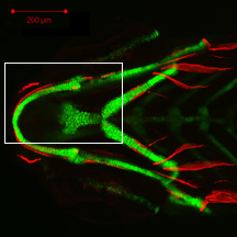
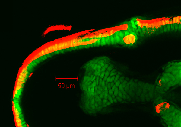
8 dpf
zc81Tg
Alizarin red
 |
|
|
| Description | By 199 hpf, Meckel’s cartilage (Mk) can has grown considerably from that seen at 149 hpf. Also visible here are the palatoquadrate (pq), dentary (d), maxilla (mx), retroarticular bone (ra), anguloarticular (aa), and quadrate (q). The dentary has now almost completely covered the lateral contour of Meckel’s cartilage in both anterior and posterior directions, including the ventrally displaced portion of Meckel’s cartilage near the mandibular symphysis (sym). The dorsal extension of the dentary can be seen to articulate with the ventral aspect of the maxilla. The anguloarticular has extended further in the perichondrium of Meckel’s cartilage both posteriorly and anteriorly, its anterior-most aspect now medial to the overlying dentary. |


 Movie(s):