FishFace
4 dpf Development
| Info |
Image
Please hold mouse over images to see annotations.
|
|
|
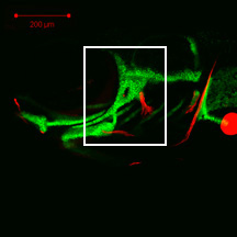
4 dpf
sp7:EGFP
Alizarin red
 |
|
|
| Description | By 96 hpf, the opercle (op) has begun to undergo a considerable shape change characterized by the addition of osteoblasts to the anterior and posterior of the ventral tip, producing a fan-shaped array of cells. The pre-existing, linear portion of the opercle has expanded slightly in width and remains flanked by osteoblasts. By this time, the most posterior branchiostegal ray 3 (bsr3) appears as a linear strut of bone surrounded by osteoblasts. The anterior tip of branchiostegal ray 3 attaches externally to the ceratohyal cartilage (not visible here). Faint EGFP-derived fluorescence throughout the image is derived from transgene expression in the ectoderm. |

 Movie(s):