
Published 30 July 2026
A new bulk RNA-seq and Cut&Run-seq dataset from the University of Southern California is now available in FaceBase!
Contributors: Lin Meng, Mingyi Zhang, Jifan Feng, Tingwei Guo, Hana Hekmat, Heliya Ziaei, Peng Chen, Aaron Harouni, Thach-Vu Ho, Yang Chai (University of Southern California)
Description:
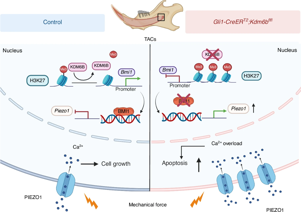
This study examined how mineralized tissues adapt to sustained mechanical stress, using mouse incisor models under varying degrees of loading. The data identify the histone demethylase KDM6B as an epigenetic regulator that preserves tissue homeostasis by protecting progenitor transit-amplifying cells from mechanical stress-induced apoptosis. Loss of Kdm6b increases H3K27me3 at the Bmi1 promoter, silencing Bmi1 and derepressing Piezo1, which drives excessive Ca2+ influx and apoptosis — while Piezo1 haploinsufficiency in Kdm6b-deficient mice rescues both the transit-amplifying cell defects and tissue homeostasis. Together the findings describe a KDM6B–H3K27me3–BMI1–PIEZO1 “mechanostat” that shields dental progenitor cells from mechanical stress. The dataset compares control and Gli1CreER;Kdm6bfl/fl adult mouse incisors by bulk RNA-seq and Cut&Run-seq.
FaceBase Dataset:
Lin Meng, Mingyi Zhang, Jifan Feng, Tingwei Guo, Hana Hekmat, Heliya Ziaei, Peng Chen, Aaron Harouni, Thach-Vu Ho, Yang Chai. Bulk RNA-seq and Cut&Run-seq analysis of control and Gli1CreER;Kdm6bfl/fl adult mouse incisors. FaceBase Consortium https://doi.org/10.25550/88-ZJCW (2026).
Publication:
Meng, L., Zhang, M., Feng, J., Guo, T., Hekmat, H., Ziaei, H., Chen, P., Harouni, A., Ho, T.-V., & Chai, Y. KDM6B safeguards mineralized tissue homeostasis from mechanical stress through epigenetic control of PIEZO1-mediated mechanotransduction in the mouse incisor. Bone Research 14(1), 59. https://doi.org/10.1038/s41413-026-00544-2 (2026).
Image: Schematic representation of KDM6B safeguarding tissue homeostasis to mechanical stress through epigenetic control of PIEZO1- mediated mechanotransduction. Using the mouse incisor as a model of mechanical loading, we reveal that within TACs, Kdm6b demethylates H3K27me3, thereby relieving the repression of the Bmi1 gene. Normal BMI1 inhibits Piezo1 expression. This maintains physiological PIEZO1 levels, ensuring calibrated Ca2+ influx for proliferation and differentiation. In contrast, loss of Kdm6b leads to an accumulation of H3K27me3 at the Bmi1 promoter region, which silences Bmi1 expression and diminishes BMI1 formation. This reduction results in pathologically increased PIEZO1 ion channels in the membrane. The subsequent Ca2+ overload triggers TAC apoptosis while reducing proliferation and differentiation. Ultimately, these molecular events compromise tissue homeostasis. Schematic created with BioRender.com. Ho, T. (2026) https://BioRender.com/8mzv4a3