FaceBase provides a unified, interoperable platform for sharing and integrating high-quality biomedical research data across biologically and systemically related domains. It supports a multitude of data types — from genomic and imaging to phenotypic data — across human studies and model organisms, enabling rigorous and reproducible science. As an exemplar of FAIR data practices and TRUST principles, FaceBase promotes standardized metadata, reliable stewardship, and AI-ready data to accelerate discovery through a unique consortium of partner resources.
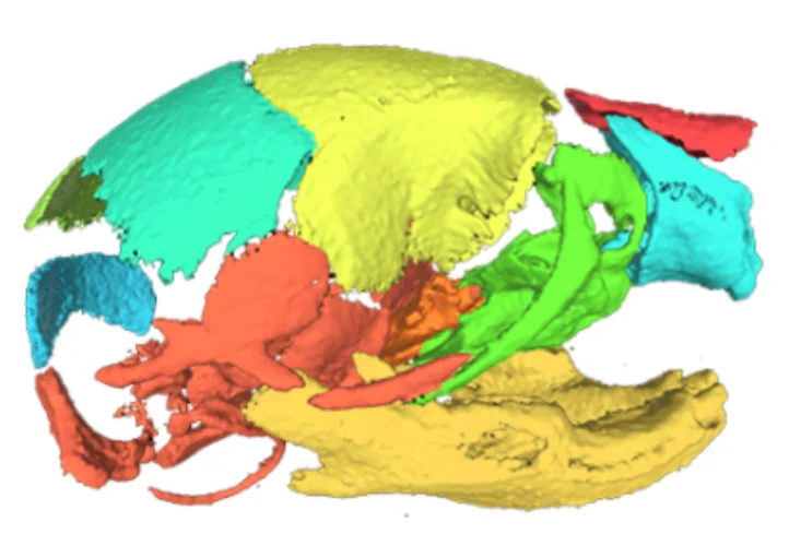
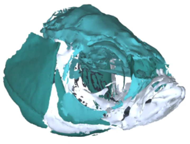
FaceBase is home to curated collections of datasets, tools, and resources organized around specific organisms and human subjects. Visit the DATA menu to explore the full range of organism-related resources.
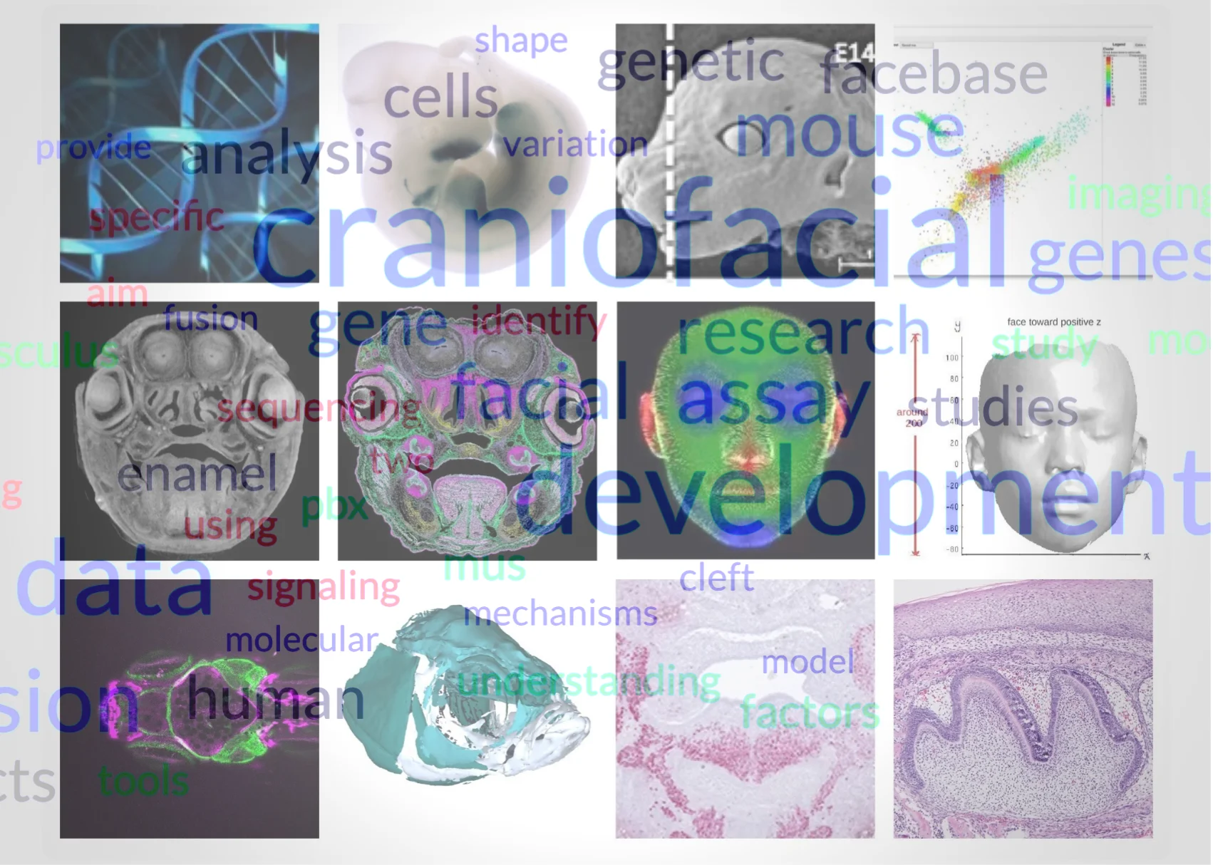
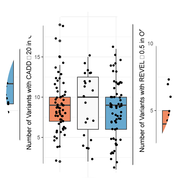
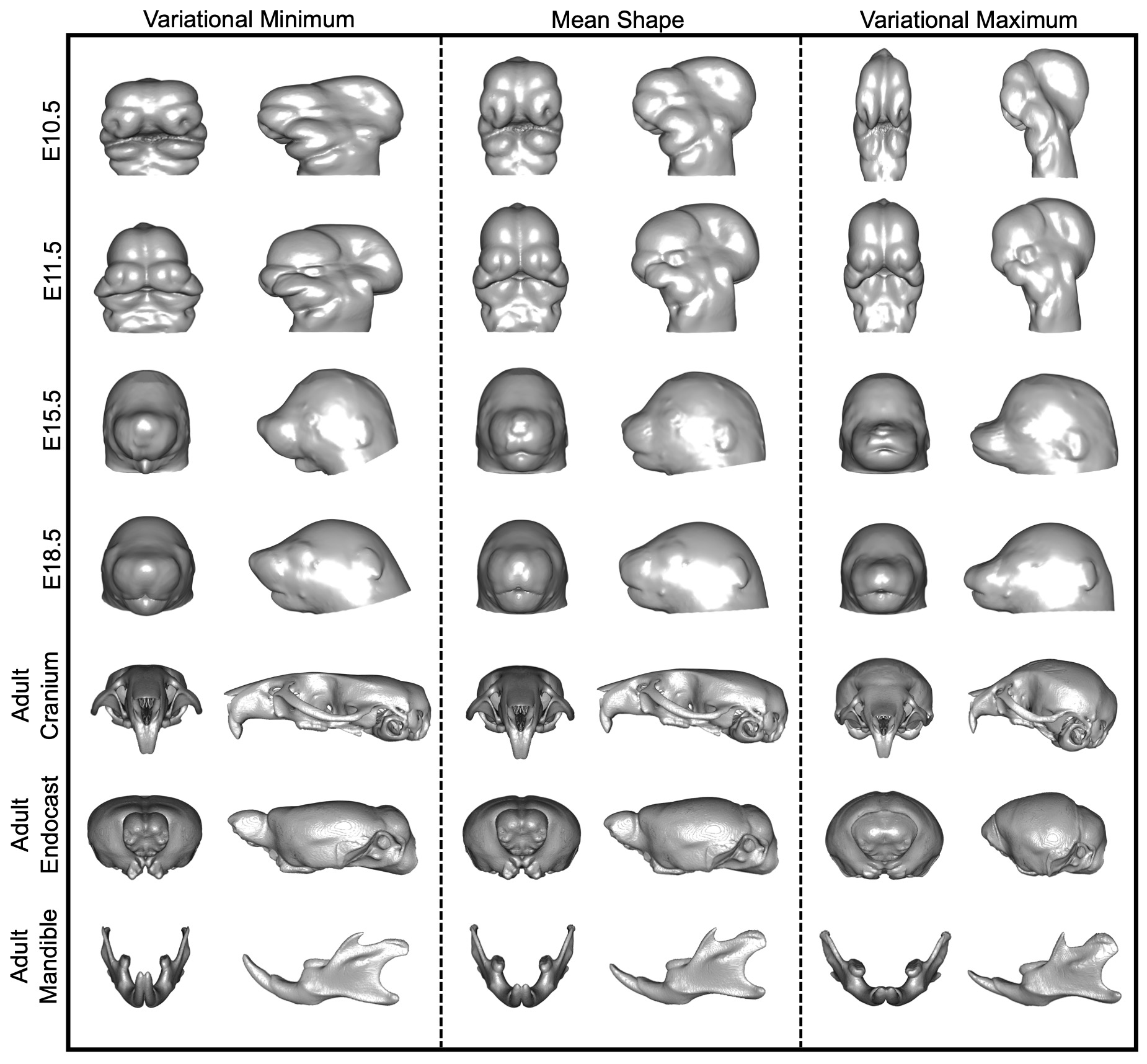
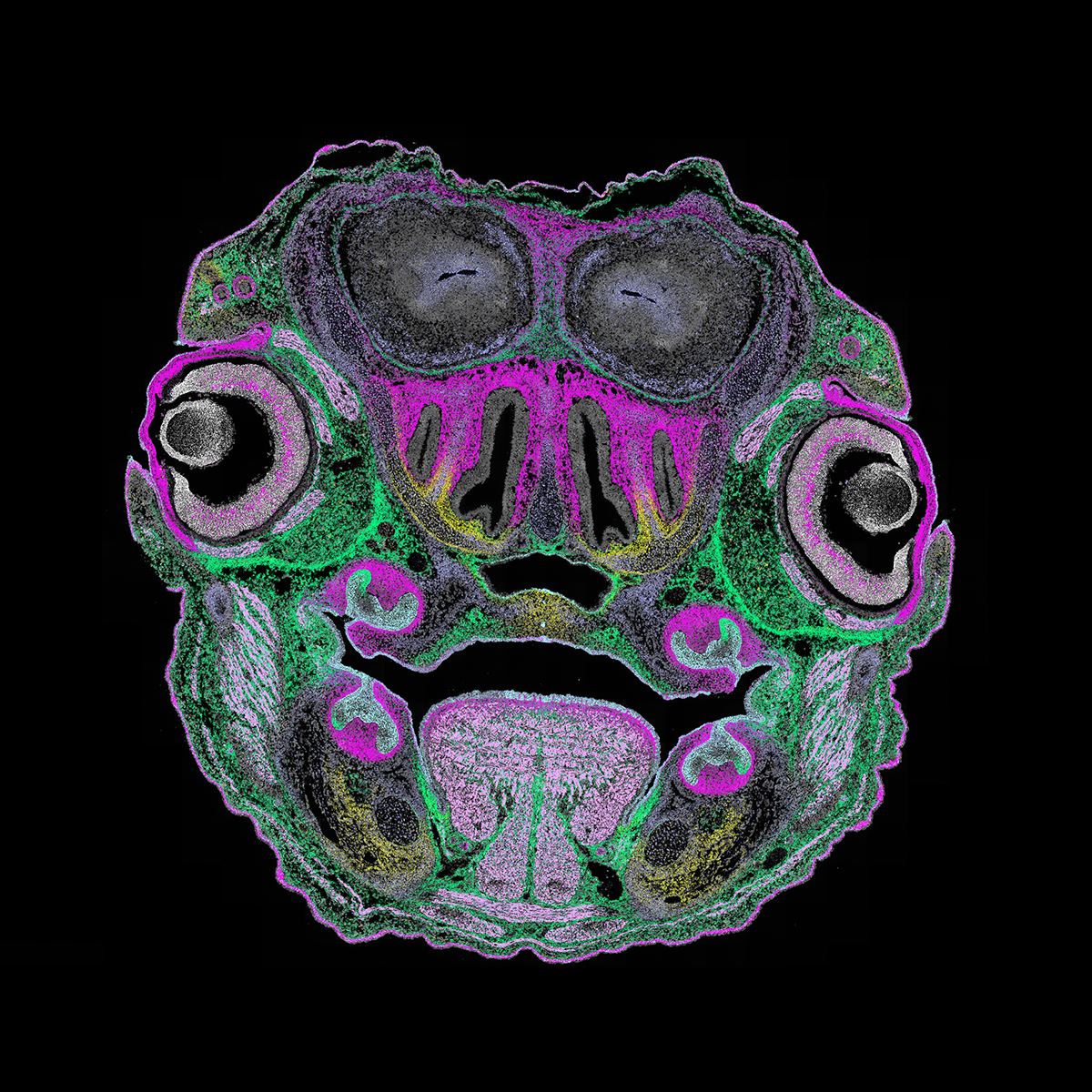
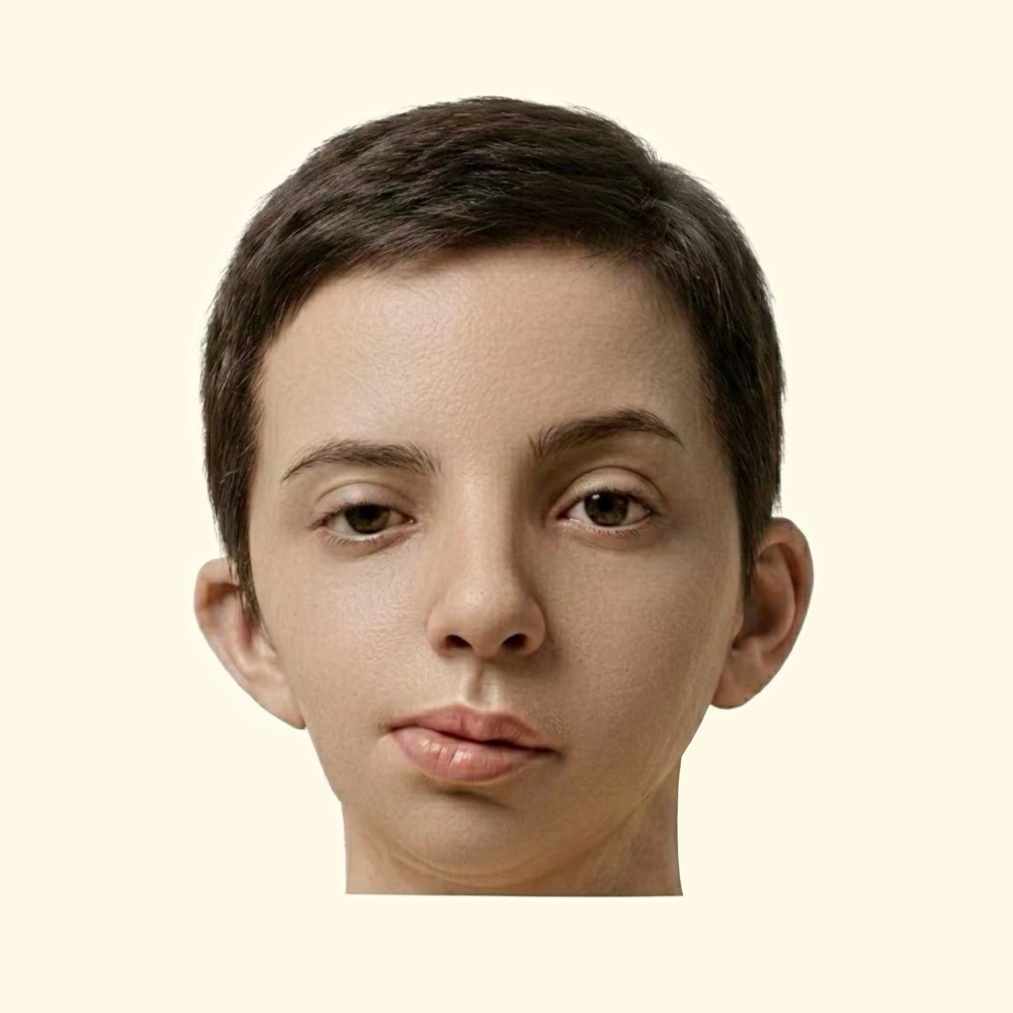
Click on the pictures to the left to learn more about these featured resources.
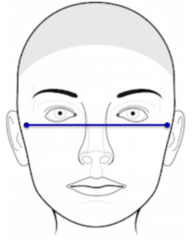
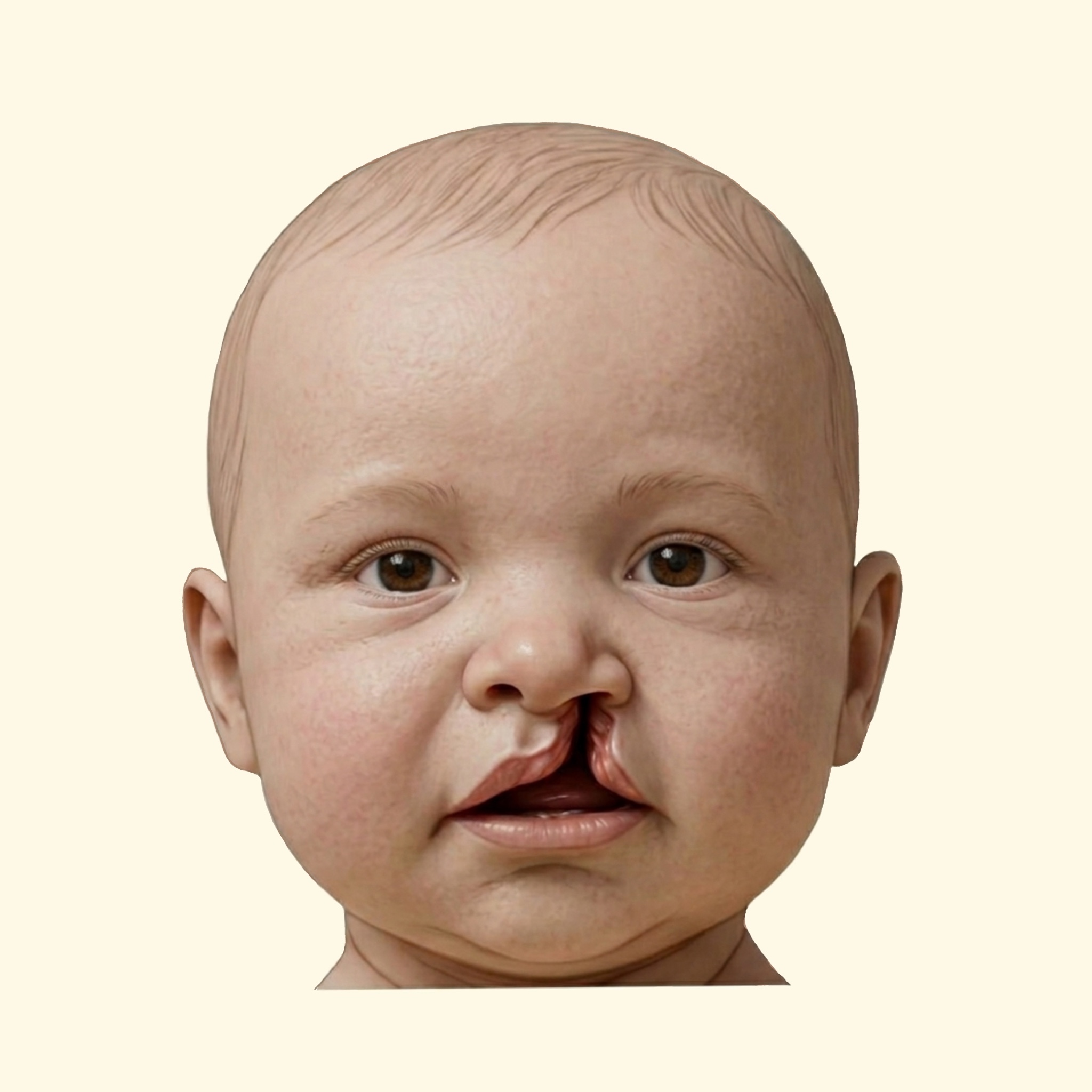
FaceBase hosts a wide assortment of disease-specific data centered on craniofacial development, dysmorphology, and related biological conditions. Visit the DATA menu to explore the full collection of disease-related resources.
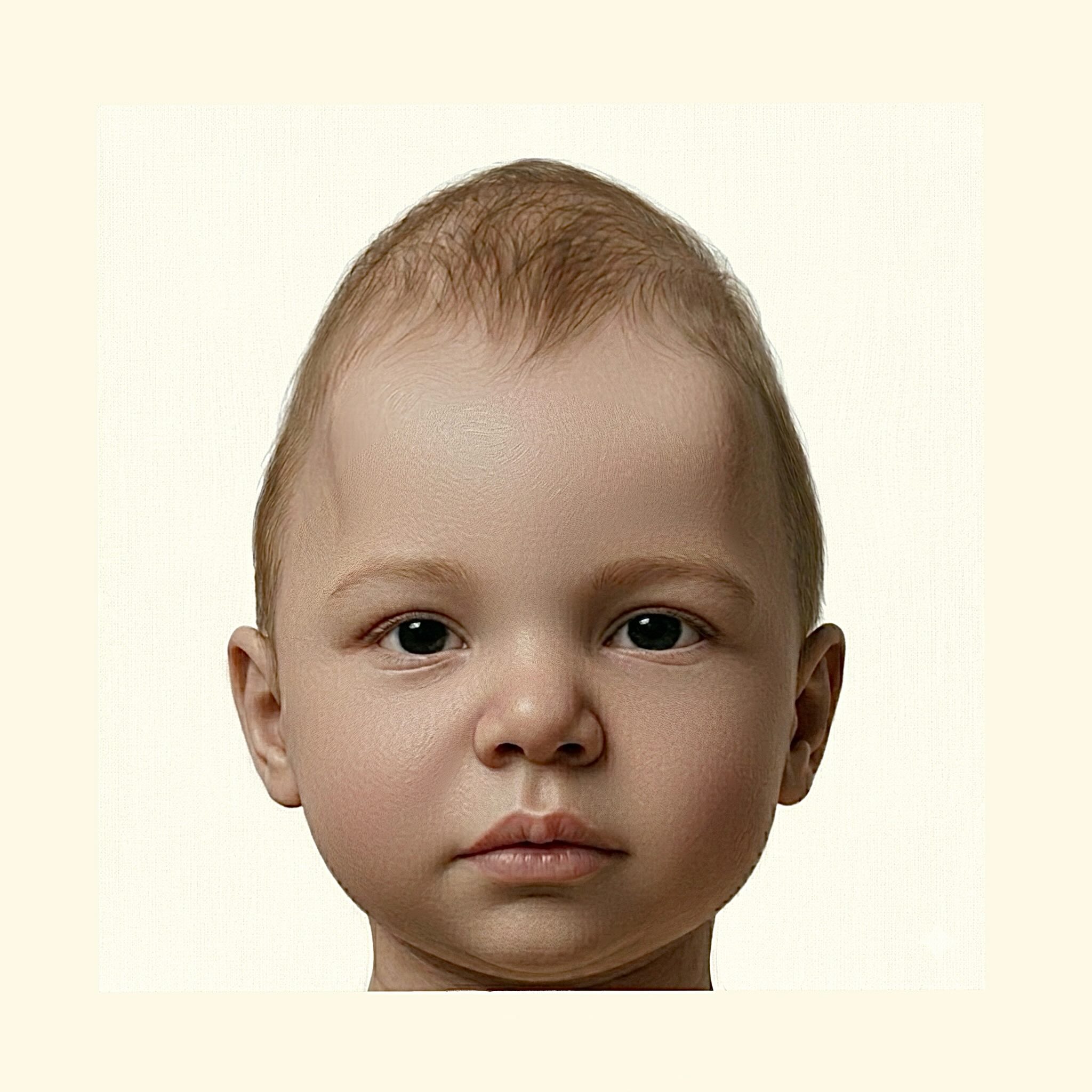
Click on the pictures to the right to learn more about these featured resources.
At the heart of FaceBase's mission is the promotion of open data sharing and collaboration guided by the FAIR principles, supported by a centralized repository that connects biologists, geneticists, clinicians, and computational scientists through interconnected datasets. This integrative ecosystem enables new hypotheses, strengthens reproducibility, and accelerates discovery across developmental biology and clinical research.
FaceBase has extensive collections of data to share with researchers worldwide. We invite you to explore .