
Published 04 August 2026
A new RNA-Seq dataset from the University of Southern California is now available in FaceBase!
Contributors: Lu Gao, Peng Chen, Jifan Feng, Tingwei Guo, Mingyi Zhang, Thach-Vu Ho, Jian-Fu Chen, Yang Chai (University of Southern California)
Description:
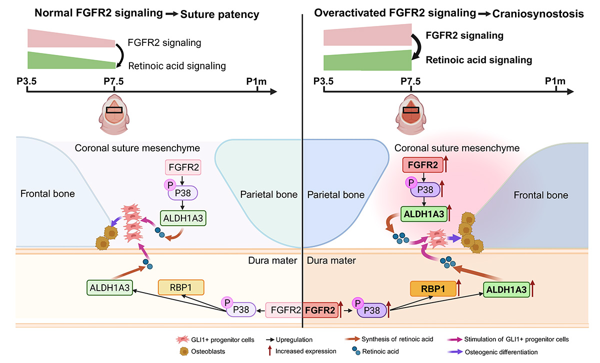
Congenital anomalies often arise during critical developmental time windows, yet the underlying mechanisms remain unclear. Here, we identify a time-specific FGFR2-retinoic acid signaling axis regulating postnatal coronal suture development through dura mater-suture mesenchyme interactions. FGFR2 signaling gradually declines after birth, leading to reduced retinoic acid signaling by moderating the expression of Aldh1a3 in the dura mater and suture mesenchyme alongside that of Rbp1 in the dura mater, thereby preserving GLI1+ progenitors and restraining osteogenesis to maintain suture patency. Importantly, FGFR2 overactivation in Fgfr2IIIc mutant mice within an early time window, disrupts this physiological FGFR2-retinoic acid signaling decline by upregulating Rbp1 and Aldh1a3 through enhanced P38 signaling. This leads to increased retinoic acid synthesis, premature osteogenic differentiation of GLI1+ progenitors, and coronal suture craniosynostosis.
Significantly, genetic restoration of retinoic acid signaling rescues craniosynostosis in Fgfr2IIIc mutant mice, confirming retinoic acid signaling as a key downstream effector of FGFR2 signaling. Notably, restoration of suture patency alone rescues neurocognitive dysfunctions in Fgfr2IIIc mutant mice despite Fgfr2 mutation in the brain, demonstrating that the neurocognitive impairments primarily arise from cranial structural constraints and elevated intracranial pressure rather than intrinsic neural defects. This critical postnatal time window in mice parallels the timing of FGFR2-related craniosynostosis onset in humans, underscoring the importance of this study in advancing our understanding of the molecular and cellular mechanisms in craniosynostosis. Our findings define a temporally regulated FGFR2-P38-retinoic acid signaling axis and highlight retinoic acid signaling as a promising therapeutic target in FGFR2 overactivation-related craniosynostosis.
FaceBase Dataset:
Lu Gao, Peng Chen, Jifan Feng, Tingwei Guo, Mingyi Zhang, Thach-Vu Ho, Jian-Fu Chen, Yang Chai. RNA-Seq elucidates the FGFR2-RA pathway in dura-suture interaction during Craniosynostosis. FaceBase Consortium https://doi.org/10.25550/8B-9CNW (2026).
Publication:
Gao, L., Chen, P., Feng, J., Guo, T., Zhang, M., Ho, T.-V., Chen, J.-F., & Chai, Y. Temporally regulated FGFR2–retinoic acid signaling mediates dura mater–suture mesenchyme interactions to prevent craniosynostosis in mice. International Journal of Oral Science. Accepted. https://sites.usc.edu/ccmb/files/2026/05/FGFR2-RA-Signaling-in-Craniosynostosis.pdf
Image: FGFR2-RA signaling governs dura mater-suture mesenchyme interaction during early postnatal development to control coronal suture development and craniosynostosis. Illustration created with BioRender.com.