
Published 01 September 2026
FaceBase has released a new dataset from the Clites Lab at UCLA that captures how the human eyelid moves and which parts of the muscle drive that motion during five distinct eyelid behaviors. It is our first dataset supported by the National Eye Institute.
Every blink spreads tear film across the cornea and clears debris from the ocular surface. Facial paralysis takes away the ability to blink, which can cause damage to the eye or infection. Efforts to restore blinking through electrical stimulation have been limited by a fundamental gap: no one has measured, at high resolution, what drives natural blink at the muscle level.
Contributors: Tyler Clites, Jinyoung Kim, Daniel Rootman (UCLA)
Description:
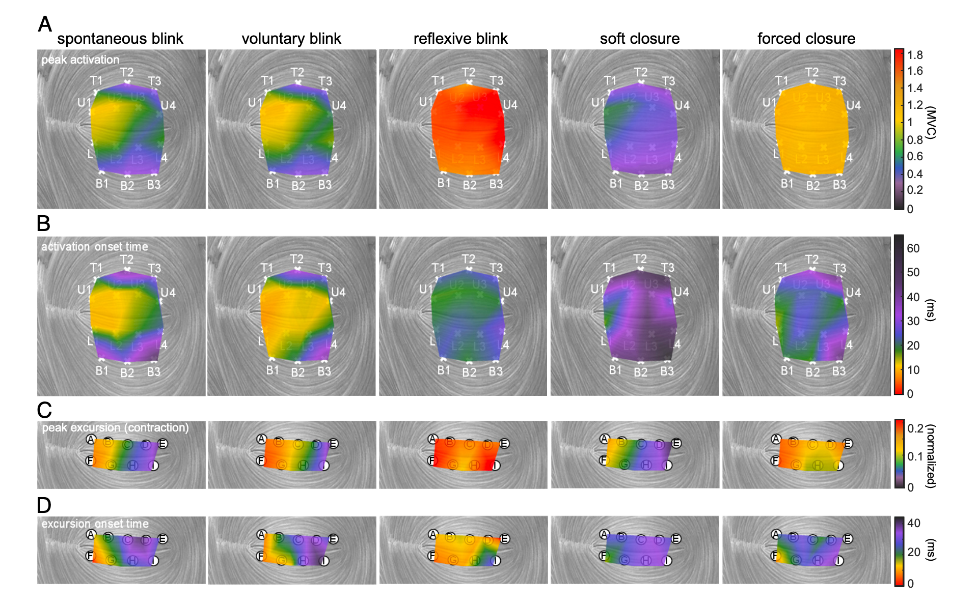
Eight adults without eyelid paralysis or other eyelid pathology (ages 22-33) performed spontaneous blink, voluntary blink, reflexive blink, soft closure, and forced closure while two systems recorded simultaneously: three-dimensional motion capture of markers along the upper and lower eyelid margins at 400 frames per second, and distributed intramuscular EMG from 14 bipolar fine-wire electrode pairs within the orbicularis oculi. The streams were hardware-time-synced, so muscle activation can be aligned frame-by-frame with the resulting eyelid motion.
The associated study, published in PNAS in 2025, found that different eyelid behaviors are produced by distinct patterns of activation across muscle segments, providing a clue as to why previous stimulation methods have been ineffective. Activating the whole eyelid at once tends to elicit a protective reflexive blink rather than the natural blink that keeps the eye wet and clear.
Supported by the National Eye Institute (award 1R21EY036680).
Accessing the data
This is restricted-access human subjects data. Researchers must complete the process described in the FaceBase data guidelines before access can be granted. Data use is limited to General Research Use (GRU) and Not-for-profit Use Only (NPU).
FaceBase Dataset:
Tyler Clites, Jinyoung Kim, Daniel Rootman. Eyelid Kinematics and Orbicularis Oculi Electromyography - Persons Without Facial Pathology. FaceBase Consortium https://doi.org/10.25550/88-28BJ (2026).
Associated Publication:
Kim J, Shirriff A, Cornwell JN, Mutis MPQ, Delis E, Wang S, Rootman DB, Clites TR. Human eyelid behavior is driven by segmental neural control of the orbicularis oculi. Proc Natl Acad Sci U S A. 2025 Aug 12;122(32):e2508058122. doi: 10.1073/pnas.2508058122. Epub 2025 Aug 7. PMID: 40773233; PMCID: PMC12358864.