Save the Date for the 2027 FaceBase Community Forum – April 27-28 in Los Angeles
July 13th, 2026
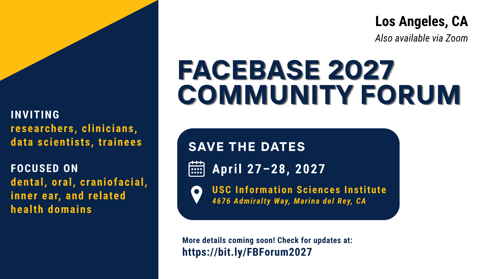
The 2027 FaceBase Community Forum will take place Tuesday, April 27 and Wednesday, April 28, 2027, in Los Angeles, CA (Marina del Rey), hosted at the USC Information Sciences Institute.
Tuesday will be a full day of programming; Wednesday will be a half day. Remote attendance via Zoom will also be available.
This gathering brings together dental, oral, and craniofacial (DOC) researchers, clinicians, and students - as well as those studying related biological systems (such as the ear and hearing research community) - working with FaceBase data and tools. An agenda, registration, and travel details will be announced in the coming months. Check back here or follow the link below for updates.
Check for updates: https://bit.ly/FBForum2027
New release: FGF signalling orchestrates multiple roles during salivary gland branching morphogenesis
May 26th, 2026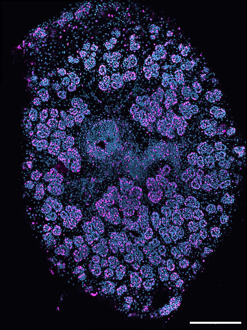
A new bulk RNA-seq dataset from King’s College London is now available in FaceBase!
Contributors: Abigail S. Tucker, Marta Perera, Joshua Brickman (King’s College London)
Description:
This study examined the role of the fibroblast growth factor (FGF) signalling pathway during branching morphogenesis in the murine embryonic submandibular salivary gland. The data compare pharmacological FGFR inhibition with conditional deletion of Fgfr2 (K14Cre;Fgfr2fl) from E13.5 +48 hours, revealing a multitude of roles for FGF signalling — including effects on fate decisions and tissue interactions. The dataset includes bulk RNA-Seq fastq files and the related counts matrix.
FaceBase Dataset:
Joshua Brickman, Abigail S. Tucker, Marta Perera. FGF signalling orchestrates multiple roles during salivary gland branching morphogenesis. FaceBase Consortium https://doi.org/10.25550/AH-J1XR (2026).
Image: Salivary gland stained with BrdU (proliferative cells, magenta) and DAPI (cyan). Image courtesy of Marta Perera.
New release: Amelogenin phosphorylation affects key regulatory genes in the enamel organ
April 30th, 2026A new amelogenin dataset is now available on FaceBase!
Contributors: Elia Beniash, Henry Margolis at the University of Pittsburgh (University of Pittsburgh)
Description:
Amelogenin (AMELX) is the predominant enamel matrix protein and has a single phosphorylation site at Serine 16 (S16), which enhances its ability to stabilize amorphous calcium phosphate in vitro. To investigate the in vivo role of AMELX phosphorylation, a knock-in mouse model (AmelxS16A) was generated in which S16 is substituted with Alanine to prevent phosphorylation. KI enamel is hypoplastic, lacks enamel rods, and features multiple ectopic calcifications; KI ameloblasts also lack Tomes’ processes and show progressive cell pathologies.
To characterize these effects comprehensively, single-cell RNA sequencing was performed on incisal enamel organs from WT and KI mice. 624 genes were differentially expressed across total enamel organ cell populations. Notably, Shh was downregulated 5.1-fold and Wnt5a was upregulated 8.1-fold in KI secretory ameloblasts compared to WT. Ten distinct cell populations were identified, with secretory ameloblasts showing the greatest transcriptomic impact, indicating that AMELX phosphorylation influences not only extracellular enamel matrix processes but also key intracellular pathways governing ameloblast biology.
FaceBase Dataset:
Elia Beniash, Henry Margolis. Amelogenin phosphorylation affects key regulatory genes in the enamel organ. FaceBase Consortium https://doi.org/10.25550/AF-V3VC (2026).
New release: Mandible vs tongue involvement in cleft palate in mouse models
April 22nd, 2026A new dataset is now available on FaceBase from Goodwin, Green, and colleagues at the University of Pittsburgh. Their study examines the respective contributions of mandibular hypoplasia and tongue malposition to cleft palate in Pierre Robin sequence (PRS), using two complementary mouse models. A related manuscript has been accepted for publication in the Journal of Dental Research.
 Comparison of palate outcomes in the Sox9fl/fl;mtHand2Cre Pierre Robin sequence model (left) and the DTA/+;mtHand2Cre micrognathia/microglossia model (right). Image courtesy of Alice Fitzgerald Goodwin.
Contributors: Alice Fitzgerald Goodwin, Jeremy Green (University of Pittsburgh)
Description:
To investigate cleft palate in Pierre Robin sequence (PRS), we generated a mouse model with Sox9 deleted specifically in the mandibular mesenchyme (Sox9fl/fl;mtHand2Cre), which resulted in mandibular hypoplasia and retrognathia, palatal shelf elevation delay, and fully penetrant cleft of the secondary palate. To determine the relative contributions of mandible vs tongue malposition to cleft palate in PRS, we generated a micrognathia and microglossia model (DTA/+;mtHand2Cre). The majority of these animals had a normally formed palate, suggesting that tongue obstruction of palatal shelf elevation is the primary contributor to cleft palate in PRS.
Data deposited on FaceBase include microCT scans of Sox9fl/fl;mtHand2Cre and control embryos at E18.5; H&E-stained coronal sections at E12.5, E13.5, E14.5, and E16.5; proliferation and apoptosis assays at E12.5 and E14.5 in the Meckel’s cartilage; immunofluorescence with antibodies against Pax7 and MHC at E14.5; RNAscope with probes against osteogenic markers at E14.5; palatal shelf explant studies at E13.5; and H&E staining and TUNEL apoptosis staining of DTA/+;mtHand2Cre and control embryos.
FaceBase Dataset:
Alice Fitzgerald Goodwin, Jeremy Green. Mandible vs tongue involvement in cleft palate in mouse models. FaceBase Consortium https://doi.org/10.25550/AB-SJQA (2026).
Publication:
Alice Fitzgerald Goodwin, Jeremy Green; Intrinsic tension drives palatal shelf reorientation post tongue retraction. Journal of Dental Research 2026. (accepted April 21, 2026 — DOI to be added upon publication)
New release: Six2 and CTCF ChIP-seq datasets of wildtype mouse embryonic facial tissues (E10.5 - E13.5)
April 16th, 2026New ChIP-seq datasets are now available in FaceBase, profiling Six2 transcription factor binding and CTCF-defined topologically associating domains in wildtype mouse embryonic facial tissues at stages E10.5 through E13.5. Input libraries are included as controls. Together these data support investigation of gene regulatory networks and chromatin architecture during craniofacial morphogenesis.
Contributors: Jingyue Xu, Han Liu and Rulang Jiang (Cincinnati Children’s Hospital, Medical Center)
Description:
We performed a ChIP-seq assay using a Six2 antibody to identify endogenous Six2-binding genomic loci in mouse embryonic facial tissues. We performed a ChIPseq assay using the CTCF antibody to identify topologically associating domains and boundaries. We generated ChIP libraries and sent them for next-generation sequencing. We also generated input libraries as control samples.
FaceBase Dataset:
Jingyue Xu, Han Liu, Rulang Jiang. Six2 and CTCF ChIP-seq datasets of wildtype mouse embryonic facial tissues (E10.5 - E13.5). FaceBase Consortium https://doi.org/10.25550/7Z-41CW (2026).