FishFace
2 dpf Development
| Info |
Image
Please hold mouse over images to see annotations.
|
|
|
2 dpf
zc81Tg
 |
|
|
| Description |
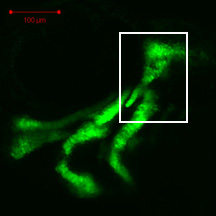
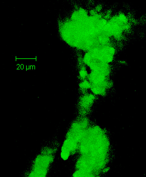
By 48 hpf, the hyosymplectic (hs) consists of a condensation of chondrogenic cells that is difficult to distinguish along its dorsal aspect from the anterior portion of the otic capsule (ot). Note the facial nerve (VII) foramen (ff) in the dorsal portion of the hyosymplectic. Ventrally, cells of the developing hyosymplectic are difficult to discern from those of the developing interhyal (ih) and ceratohyal (ch), whereas there is clear separation from cells of the more anterior developing palatoquadrate (pq). No Alcian blue staining of matrix is observed in these developing cartilages at this stage (data not shown). The hyosymplectic can be separated into dorsal (i.e., hyomandibula) and ventral (i.e., symplectic) regions, which at this timepoint are divided by a distinctive area of decreased fluorescence (*).
* See image at https://fishstix.uoregon.edu/kimmel-atlas/Hyosymplectic.html |
 Movie(s):