FishFace
The palatoquadrate - associated bones and articulations
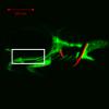
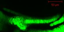
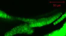
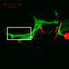
This series of images from late embryonic stages--2 days post-fertilization (dpf)--to late larval stages--21dpf--shows the appearance, overall anatomical arrangements, and gross morphology of cartilage and bone elements of the dorsal jaw skeleton (Arch 1) of the developing zebrafish craniofacial apparatus. We use the zc81Tg transgenic zebrafish, with fluorescent chondrocytes, to visualize cartilage and Alizarin red to visualize bone. Images are taken from both ventral (with lateral to top) and lateral views.
|
Info
Click linked text in this column to see full page image(s)
|
Image
Please hold mouse over images to see annotations.
Click on magnifying icon to view large image. |
Description
Click on an underlined term to see its definition and link to related pages
|
|
|
zc81Tg
|
|
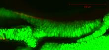
By 51 hpf, the palatoquadrate (pq) consists of a condensation of chondrogenic cells that is difficult to distinguish along its anterior aspect from the posterior portion of Meckel’s cartilage (Mk). Note the dimmer GFP signal in the region (*) between the palatoquadrate and Meckel’s cartilage. No Alcian blue staining of matrix is observed in these developing cartilages at this stage (data not shown). |
|
|
zc81Tg
Alizarin red
|
|
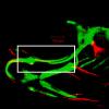
By 72 hpf, the palatoquadrate (pq) has increased in size and cell number from that seen at 51 hpf. The palatoquadrate now can be discerned more easily from Meckel’s cartilage (Mk) in its anterior articulation and articulates considerably with the hyosymplectic (hs) posteriorly. Alcian blue staining of matrix is observed in these developing cartilages at this stage (data not shown). The pterygoid process (ptp) of the palatoquadrate can be seen growing dorsomedially from the main body to approximate the ethmoid (e) of the neurocranium. A more ventral view demonstrates that, similar to the pterygoid process, the main body of the palatoquadrate is approximately one cell layer thick. |
|
|
zc81Tg
Alizarin red
|
|
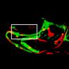
By 104 hpf, the palatoquadrate (pq) has grown considerably from that seen at 72 hpf. The palatoquadrate now can be discerned completely from Meckel’s cartilage (Mk) at its anterior articulation and articulates with the hyosymplectic (hs) posteriorly. The pterygoid process (ptp) of the palatoquadrate articulates with the ethmoid (e) of the neurocranium. Also visible is the entopterygoid (en), which initiates in the dermis alongside the medial aspect of the pterygoid process. |
|
|
zc81Tg
Alizarin red
|
|
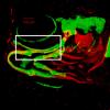
By 146 hpf, the palatoquadrate (pq) has grown considerably from that seen at 104 hpf. The palatoquadrate articulates with Meckel’s cartilage (Mk) anteriorly, the hyosymplectic (hs) posteriorly, and the ethmoid (e) of the neurocranium through its pterygoid process (ptp) dorsomedially. Palatoquadrate chondrocytes remain aligned in a single row of cells, visible in ventral view. A partial projection in the ventral view demonstrates that the entopterygoid (en) is medial to, and spans 2/3 of the anterior-posterior length of, the palatoquadrate. The quadrate (q) is now visible in the lateral aspect of the antero-ventral perichondrium of the palatoquadrate near its articulation with Meckel’s cartilage and extends from the perichondrium into the dermis ventral to the main body of the palatoquadrate. |
|
|
zc81Tg
Alizarin red
|
|
By 200 hpf, the palatoquadrate (pq) has grown from that seen at 146 hpf, but retains the same overall shape. The palatoquadrate articulates with Meckel’s cartilage (Mk) anteriorly and the hyosymplectic (hs) posteriorly. The ventral view demonstrates that the entopterygoid (en) forms medially along 2/3 the anterior-posterior length of the palatoquadrate, including the pterygoid process (ptp). The quadrate (q) is visible in both the lateral and medial perichondrium of the palatoquadrate near the articulation with Meckel’s and has begun to extend dorsally. The quadrate also extends posteriorly outside the perichondrium into the dermis lateral to the hyosymplectic.
|
|
|
zc81Tg
Alizarin red
|
|
By 242 hpf, the palatoquadrate (pq) increased in size from that seen at 200 hpf, but retains its earlier shape. The palatoquadrate articulates with Meckel’s cartilage (Mk) anteriorly and the hyosymplectic (hs) posteriorly. The entopterygoid (en) grows medially along 2/3 the anterior-posterior length of the palatoquadrate, including the pterygoid process (ptp). The quadrate (q) continues to extend dorsally and medially within the perichondrium of the palatoquadrate near the articulation with Meckel’s cartilage and also continues to extend posteriorly into the dermis lateral to the symplectic (sy) in the perichondrium of the hyosymplectic. |
|
|
zc81Tg
Alizarin red
|
|
By 339 hpf, the palatoquadrate (pq) has grown from that seen at 242 hpf, retaining its earlier shape. The palatoquadrate articulates with Meckel’s cartilage (Mk) anteriorly and the hyosymplectic (hs) posteriorly. The entopterygoid (en) is medial along 2/3 the anterior-posterior length of the palatoquadrate, including the pterygoid process (ptp). The portion of the quadrate (q) in the perichondrium of the palatoquadrate near the articulation with Meckel’s cartilage has expanded dorsally and posteriorly into a fan shape. The dermal portion of the quadrate continues to extend posteriorly, covering the symplectic (sy) laterally. The metapterygoid (mpt) can be seen in the perichondrium of the posterior portion of the main body of the palatoquadrate. |
|
|
zc81Tg
Alizarin red
|
|
By 510 hpf, the palatoquadrate (pq) has grown considerably from that seen at 339 hpf, but its overall shape has remained basically the same since that achieved at 146 hpf. The palatoquadrate articulates with Meckel’s cartilage (Mk) anteriorly and the hyosymplectic (hs) posteriorly. The quadrate (q) has now spread through the perichondrium to cover part of the anterior portion of the palatoquadrate, excluding the pterygoid process (ptp). The ectopterygoid (ect) can be seen in the dermis running antero-ventral to the lateral portion of the pterygoid process of the palatoquadrate, approaching the Meckel’s-palatoquadrate joint. The metapterygoid (mpt) has spread in the perichondrium of the posterior portion of the main body of the palatoquadrate. Also visible is the scleral (sc) cartilage of the eye. |


 Movie(s):