FishFace
2.5 dpf Development
| Info |
Image
Please hold mouse over images to see annotations.
|
|
|
2.5 dpf
zc81Tg
 |
|
|
| Description |
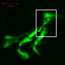
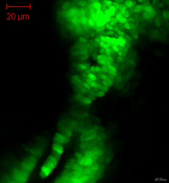
By 55 hpf, cellular boundaries of chondrocytes of the hyosymplectic (hs) are more easy to distinguish, although cells of the developing hyosymplectic remain difficult to discern from those of the developing otic capsule (ot), interhyal (ih) and ceratohyal (ch). The more dorsal hyomandibula and the more ventral symplectic regions of the hyosymplectic are still separated by a distinctive area of decreased fluorescence (*). Alcian blue staining of matrix is observed in these developing cartilages at this stage (data not shown). |
 Movie(s):