FishFace
3 dpf Development
| Info |
Image
Please hold mouse over images to see annotations.
|
|
|
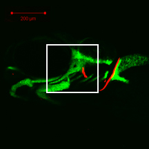
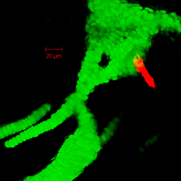
3 dpf
zc81Tg
Alizarin red
 |
|
|
| Description |
By 72 hpf, the hyosymplectic (hs) has increased in size and cell number from that seen at 48 hpf. The hyomandibula region of the dorsal hyosymplectic is still difficult to distinguish from the anterior portion of the otic capsule (ot) dorsally, but the symplectic region of the ventral hyosymplectic can be discerned easily from the ceratohyal (ch) ventrally. The interhyal (ih), which articulates where the two hyosymplectic regions meet, is still not easy to distinguish. The symplectic region overlaps considerably with the palatoquadrate (pq) anteriorly. Note the facial nerve (VII) foramen (ff) in the hyomandibula region. Alcian blue staining of matrix is observed in these developing cartilages at this stage (data not shown). The opercle (op) projects ventrally from the posterodorsal aspect of the hyomandibula region in a ball-and-socket joint. |
 Movie(s):