FishFace
14 dpf Development
| Info |
Image
Please hold mouse over images to see annotations.
|
|
|
14 dpf
zc81Tg
Alizarin red
 |
|
|
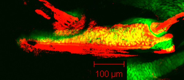
| Description | By 338 hpf, Meckel’s cartilage (Mk) and the palatoquadrate (pq), dentary (d), premaxilla (pm), maxilla (mx), retroarticular bone (ra), anguloarticular (aa), and quadrate (q) are visible. The dentary has now covered the circumference of Meckel’s cartilage near the mandibular symphysis (sym), which is unlike that seen along the rest of the length of Meckel’s cartilage, where it only extends along one edge (i.e., laterally). Meckel’s cartilage now is displaced dorsally at the mandibular symphysis (data not shown). Note the extensive articulation between the dentary and maxilla, as well as the developiing articulation between the anguloarticular, quadrate, and retroarticular in the jaw joint. |


 Movie(s):