FishFace
6dpf Development
| Info |
Image
Please hold mouse over images to see annotations.
|
|
|
6dpf
zc81Tg
Alizarin red
 |
|
|
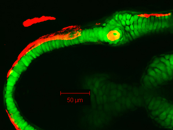
| Description | By 149 hpf, Meckel’s cartilage (Mk) has grown in size from that seen at 105 hpf. Note the appearance of chondrocyte doublets along the body of the Meckel’s cartilage in the lateral view, which is consistent with the idea that growth along the length of the cartilage is also accomplished by division of resident chondrocytes. Also visible in these images are the palatoquadrate (pq), dentary (d), maxilla (mx), retroarticular bone (ra), anguloarticular (aa), and quadrate (q). The dentary follows the lateral contour of Meckel’s in both anterior and posterior directions, projecting away from the cartilage along its posteroventral aspect. The anguloarticular appears in the posterolateral perichondrium of Meckel’s cartilage. Cells near the mandibular symphysis (sym) are positioned ventrally relative to the trajectory of the main body of Meckel’s cartilage. |


 Movie(s):