FishFace
60 hpf Development
[ Back to Pharyngeal development: Pre-skeletal]
| Info | Image Please hold mouse over images to see annotations. |
|
|
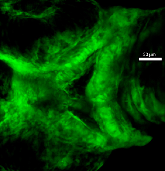
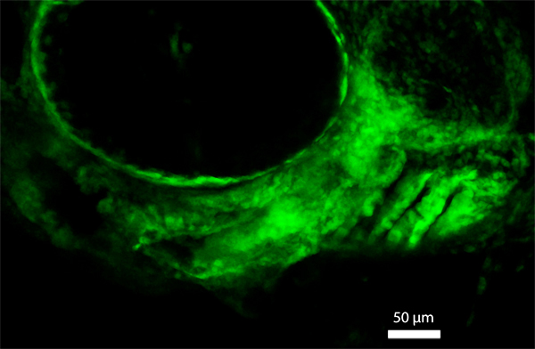
60 hpf
fli1a:EGFP
|
|
|
| Description |
By 60 hpf, neural crest-derived cells have begun to condense into distinct skeletal components of the craniofacial apparatus (see Pharyngeal arch development--Skeletal elements). In fact, Alcian blue staining of cartilage matrix is visible in skeletal elements of pharyngeal arches 1 and 2 at this timepoint (data not shown). Cells of Meckel’s cartilage (Mk) and the palatoquadrate (pq) can be discerned in pharyngeal arch 1, while cells of the hyosymplectic (hs) and the ceratohyal (ch) are visible in pharyngeal arch 2. The ventral region of pharyngeal arch 2 has expanded extensively from that seen at 48 hpf, so that cells from the left and right sides of the embryo meet at the midline between the developing ceratohyals. |