EnamelBase: A Primer on Amelogenesis
Enamel formation (amelogenesis) involves a number of epithelium-derived cell types. The innermost layer, the inner enamel epithelium, is a single layer of cells that differentiate into ameloblasts. The outermost layer is also a single layer of cells, referred to as the outer enamel epithelium. The star-shaped cells of stellate reticulum and the stratum intermedium are connected to each other and to the outer and inner enamel epithelium, respectively. The inner and outer enamel epithelium converge at a region called the cervical loop, which is a niche for epithelial stem cells and thus provides a constant source of enamel-forming cells until the enamel is matured and the tooth crown is fully formed but with one exception; that is, in rodent incisors the stem cell niche in the cervical loop is retained for life enabling the continuous growth of these teeth.
The cells comprising the enamel organ in the cervical loop, secretory stage, and maturation stage are morphologically distinct. Function of the cervical loop and activities of the stem cells remain partially defined. Secretory and maturation stages are better understood. Studies have clearly illustrated the changing ameloblast morphologies throughout amelogenesis as viewed histologically (See the figure below).
Secretary stage: During the secretory stage, four cell populations are easily recognized in the developing tooth organ: a single layer of secretory ameloblasts; the stratum intermedium, typically one or two cell layers thick; the stellate reticulum comprised of a larger grouping of star-shaped cells; and the single-layer outer enamel epithelium.
Maturation stage: The anatomy of the enamel organ changes quickly and dramatically from secretory to maturation stage. After a brief transition period, secretory ameloblasts become shorter; they have frequently been referred to as squatter maturation cells. The other three epithelial cell populations identified in the secretory stage (stratum intermedium, stellate reticulum and outer enamel epithelium) reorganize to become the papillary layer (PL) in juxtaposition to the maturation ameloblasts and are rich with blood vasculature weaving through its folds.
With the financial support of the National Institute of Dental and Craniofacial Research (NIDCR) through UH2/UH3 Cooperative Agreements awards, four enamel-focused research groups have developed a number of cultured cell and animal models to study amelogenesis, and have also developed methodologies to investigate, at the nanoscale level, enamel atomic composition of developing and mature enamel. These four groups are headed by Thomas Diekwisch (University of Rochester Medical Center); Jan Hu (University of Michigan); Derk Joester (Northwestern University); Ophir Klein (Cedars-Sinai Medical Center, Los Angeles); and Michael Paine (University of Southern California).
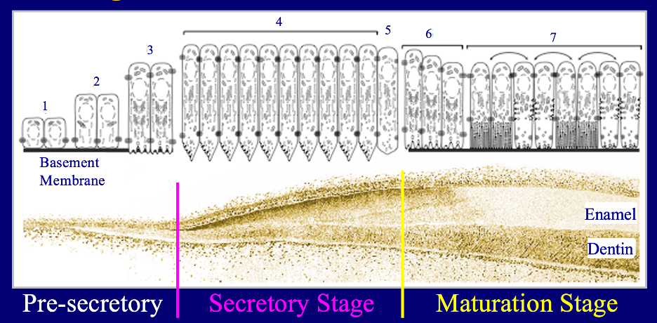
Figure: Stages of Dental Enamel Formation This image shows the stages of enamel formation and how ameloblasts change through the process. Enamel formation can be divided into 3 stages: presecretory, secretory, and maturation stages. On top is a rendering of 7 ameloblast morphologies associated with the progression of enamel formation in the histology of a developing tooth below. The presecretory ameloblasts, guided by signals from the underlying ectomesenchyme, divide to establish a sheet of cells, grow taller, polarize (nucleus toward the top), expand their secretory apparatus, add proximal tight junctions, penetrate, and resorb the basement membrane. Secretory ameloblasts form the initial enamel ribbons on the surface of freshly mineralized collagen on the dentin surface and then grow a specialized distal surface (from the terminal bars/tight junctions to the tip) called a Tomes’ process, which has separate secretory zones that extend enamel ribbons in different directions that established the rod/interrod organization characteristic of mammals. The enamel ribbons grow predominantly in length and lay out the entire thickness of the enamel layer. Maturation ameloblasts modulate between ruffle-ended and smooth-end phases to remove residual protein from between the thin crystallites, allowing them to widen into each other and harden (enamel maturation).