FishFace
4 dpf Development
[ Back to Pharyngeal development: Pre-skeletal]
| Info | Image Please hold mouse over images to see annotations. |
|
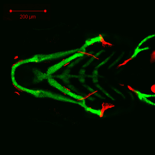
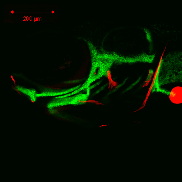
4 dpf
zc81Tg
Alizarin red
|
Ventral

Lateral

 Movie(s): Movie(s):
4 dpf ventral overview (file missing) Movie(s):
|
| Description |
By 105 hpf, this decreased magnification view shows the basic anatomical arrangement of the entire early zebrafish pharyngeal skeleton, including Meckel’s cartilage (Mk), palatoquadrate (pq), basihyal (bh), ceratohyal (ch), interhyal (ih), hyosymplectic (c) (hs), copula 1, and hypobranchial cartilages and the ceratobranchial bone (cbb). Alizarin red staining indicates the opercle (op) and ceratobranchial 5 (cb5) bones, as well as the appearance of the maxilla (mx), dentary (d), and retroarticular (ra) bones, derived from the first pharyngeal arch, and ceratohyal bone (chb), branchiostegal ray 3 (bsr3), and hyomandibula (hm) bones, derived from the second pharyngeal arch, during the fourth day of development. Also visible are the cleithrum (cl) bone and the coraco-scapular (cor) cartilage of the pectoral fin skeleton. |