FishFace
6 dpf Development
| Info |
Image
Please hold mouse over images to see annotations.
|
|
|
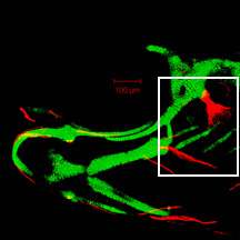
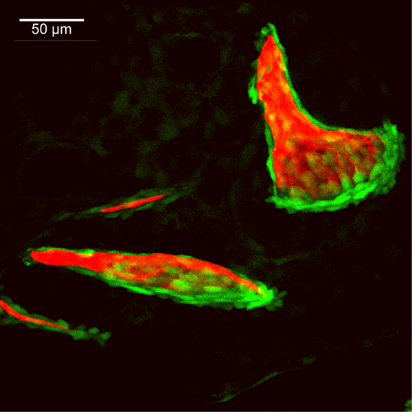
6 dpf
sp7:EGFP
Alizarin red
 |
|
|
| Description | By 144 hpf, the ventral edge of the opercle (op) has continued its major mode of bone growth both ventrally and posteriorly, resulting in a considerable size increase from that seen at 120 hpf. Branchiostegal ray 3 (bsr3), no longer rod-shaped, continues to grow along its posteroventral edge, with its future shape and size predicted by the arrangement of osteoblasts. The interopercle (iop) has added more osteoblasts and increased its linear growth from that seen at 120 hpf. The more anterior and ventral branchiostegal ray 2 (bsr2) is now apparent. Faint EGFP-derived fluorescence throughout the image is derived from transgene expression in the ectoderm. The hyomandibula is not visible in this image. |
 Movie(s):