FishFace
Evidence for a zebrafish mentomeckelian bone
Cellular resolution of bones covering anterior Meckel’s cartilage suggests that a zebrafish chondral mentomeckelian bone is fused to the dermal dentary, as seen in other teleosts. First, we observed only one condensation of osteoblasts in the anterior portion of the developing mandible at 3 dpf. Second, confocal slices of Alizarin-red stained larvae at 8 dpf demonstrate a rather continuous mineralized bone matrix extending from near the mandibular symphysis to include the dentary more postero-laterally. The anterior region of bone matrix, which we interpret as the mentomeckelian, intimately covers chondrocytes in the fashion of a chondral bone, whereas the dentary more posteriorly is well-separated from the cartilage, over a layer we take to be the perichondrium. The latter arrangement is typical of dermal bone.
|
Info
Click linked text in this column to see full page image(s)
|
Image
Please hold mouse over images to see annotations.
Click on magnifying icon to view large image. |
Description
Click on an underlined term to see its definition and link to related pages
|
|
|
sp7:EGFP
Alizarin red
|
|
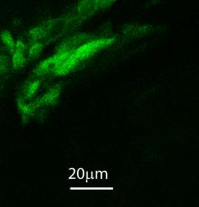
Before bone matrix is detected by Alizarin red, only one group of osteoblasts is apparent by 78 hpf in the anterior mandible of Alizarin red-stained sp7:EGFP larvae. This condensation may represent osteoblasts that will form both the mentomeckelian (mm) anteriorly and the dentary (d) posteriorly. Expression of the transgene is also visible in ectodermal cells (ecto) of the jaw epithelium.
|
|
|
fli1a:EGFP
Alizarin red
|
|
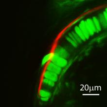
From a ventral view, confocal slices of the anterior region of Meckel’s cartilage (Mk) in 8 dpf Alizarin red-stained fli1a:EGFP larvae show that the bone matrix of the presumptive mentomeckelian (mm) intimately overlies chondrocytes anteriorly (yellow arrow). More posteriorly, a thin fli1a:EGFP expressing layer, which we interpret as the Meckel’s cartilage perichondrium (magenta arrow), intervenes between the dentary (d) and chondrocytes of Meckel’s cartilage.
|


 Movie(s):
