FishFace
10 dpf Development
| Info |
Image
Please hold mouse over images to see annotations.
|
|
|
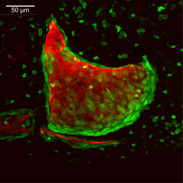
10 dpf
sp7:EGFP
Alizarin red
 |
|
|
| Description | By 240 hpf, osteoblasts line the anterior edge of the opercle (op) as an additional growth mode begins to extend this edge anteriorly. Continued posteroventral outgrowth of the opercle adds substantial size to that seen at 192 hpf. The interopercle (iop) has expanded dorsal-ventrally along its posterior end as it grows toward the opercle. Ossification of the rod-shaped subopercle (sop) has initiated just ventral and medial to the opercle. Osteoblast populations of the opercle and subopercle are separated from one another by several cell layers of mesenchyme (data not shown). The branchiostegal rays are not visible in this particular plane of view. Bone matrix of the hyomandibula (hm) can be seen in the perichondrium of the hyosymplectic (not visible here), although the flattened, small osteoblasts of the hyomandibula are not seen at this magnification. Foci of EGFP-derived fluorescence across the image are derived from transgene expression in the ectoderm. |
 Movie(s):