FishFace
The opercle - associated bones and articulations
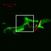
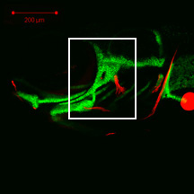
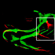
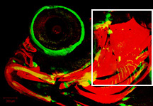
This series of images from late embryonic stages--3 days post-fertilization (dpf)--to late larval stages—21 dpf--shows the appearance, overall anatomical arrangements, and gross morphology of bone elements of the dorsal jaw-supporting skeleton (Arch 2) of the developing zebrafish craniofacial apparatus. We use both sp7:EGFP transgenic zebrafish, with fluorescent osteoblasts, and Alizarin red to visualize bone, and all images here are taken from lateral view.
|
Info
Click linked text in this column to see full page image(s)
|
Image
Please hold mouse over images to see annotations.
Click on magnifying icon to view large image. |
Description
Click on an underlined term to see its definition and link to related pages
|
|
|
sp7:EGFP
Alizarin red
|
|
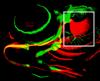
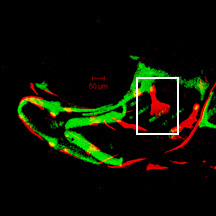
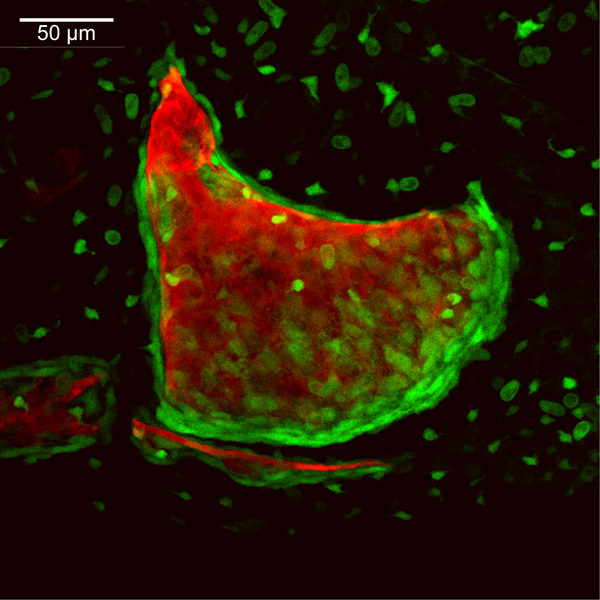
By 72 hpf, the opercle (op) consists of a linear spicule of bone matrix surrounded by tightly packed osteoblasts. The opercle articulates along its dorsal tip with the hyosymplectic cartilage (not visible here) through a ball and socket joint. At this time, opercle growth occurs ventrally through the addition of osteoblasts in a linear fashion that parallels the existing bone (data not shown). Faint EGFP-derived fluorescence throughout the image is derived from transgene expression in the ectoderm.
|
|
|
sp7:EGFP
Alizarin red
|
|
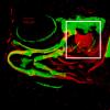
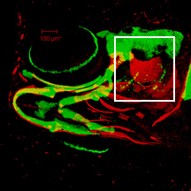
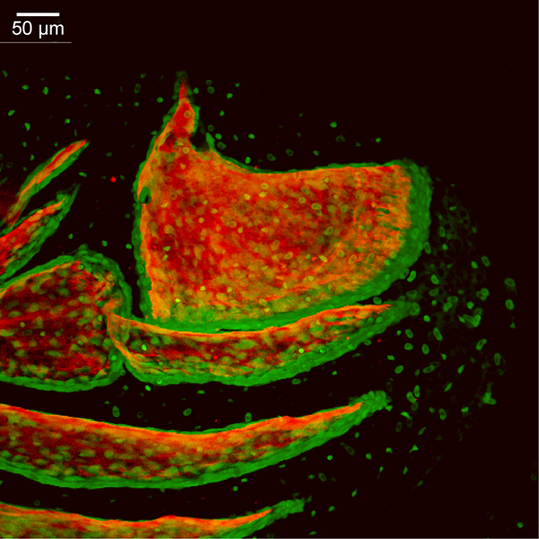
By 96 hpf, the opercle (op) has begun to undergo a considerable shape change characterized by the addition of osteoblasts to the anterior and posterior of the ventral tip, producing a fan-shaped array of cells. The pre-existing, linear portion of the opercle has expanded slightly in width and remains flanked by osteoblasts. By this time, the most posterior branchiostegal ray 3 (bsr3) appears as a linear strut of bone surrounded by osteoblasts. The anterior tip of branchiostegal ray 3 attaches externally to the ceratohyal cartilage (not visible here). Faint EGFP-derived fluorescence throughout the image is derived from transgene expression in the ectoderm.
|
|
|
sp7:EGFP
Alizarin red
|
|
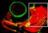
At 120 hpf, the opercle (op) is now fan-shaped, with a high density of osteoblasts along the fast-growing ventral edge. A single layer of osteoblasts outlines the other, more slowly growing edges of the bone. Branchiostegal ray 3 (bsr3) has grown posteriorly away from its articulation with the ceratohyal (not visible here), and is lined by osteoblasts along its posteroventral edge. Initial ossification and osteoblasts of the interopercle (iop) are apparent at this time between the branchiostegal ray 3 and opercle. Bone matrix of the hyomandibula (hm) can be seen in the perichondrium of the hyosymplectic (not visible here), although the flattened, small osteoblasts of the hyomandibula are not seen at this magnification. Faint EGFP-derived fluorescence throughout the image is derived from transgene expression in the ectoderm
|
|
|
sp7:EGFP
Alizarin red
|
|
By 144 hpf, the ventral edge of the opercle (op) has continued its major mode of bone growth both ventrally and posteriorly, resulting in a considerable size increase from that seen at 120 hpf. Branchiostegal ray 3 (bsr3), no longer rod-shaped, continues to grow along its posteroventral edge, with its future shape and size predicted by the arrangement of osteoblasts. The interopercle (iop) has added more osteoblasts and increased its linear growth from that seen at 120 hpf. The more anterior and ventral branchiostegal ray 2 (bsr2) is now apparent. Faint EGFP-derived fluorescence throughout the image is derived from transgene expression in the ectoderm. The hyomandibula is not visible in this image.
|
|
|
sp7:EGFP
Alizarin red
|
|
By 168 hpf, the ventral edge of the opercle (op) has continued its major mode of bone growth both ventrally and posteriorly, increasing in size from that seen at 144 hpf. Branchiostegal ray 3 (bsr3) continues to grow along its posteroventral edge. The interopercle (iop) has extended both anteriorly and posteriorly but remains a simple spicule of bone surrounded by a thin layer of osteoblasts. Likewise, branchiostegal ray 2 (bsr2) has remained rod-shaped, growing posteriorly, where numerous osteoblasts are visible. Bone matrix of the hyomandibula (hm) can be seen in the perichondrium of the hyosymplectic (not visible here), although the flattened, small osteoblasts of the hyomandibula are not seen at this magnification. Faint EGFP-derived fluorescence throughout the image is derived from transgene expression in the ectoderm.
|
|
|
sp7:EGFP
Alizarin red
|
|
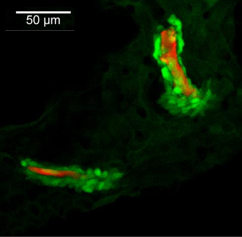
By 192 hpf, continued posteroventral expansion of the opercle (op) and branchiostegal ray 3 (bsr3) results in overall size increases in these bones relative to that seen at 168 hpf. The interopercle (iop) has grown in dorsal-ventral width and has more osteoblasts along its edges than seen at 168 hpf. Bone matrix of the hyomandibula (hm) can be seen in the perichondrium of the hyosymplectic (not visible here), although the flattened, small osteoblasts of the hyomandibula are not seen at this magnification. Foci of EGFP-derived fluorescence across the image are derived from transgene expression in the ectoderm.
|
|
|
sp7:EGFP
Alizarin red
|
|
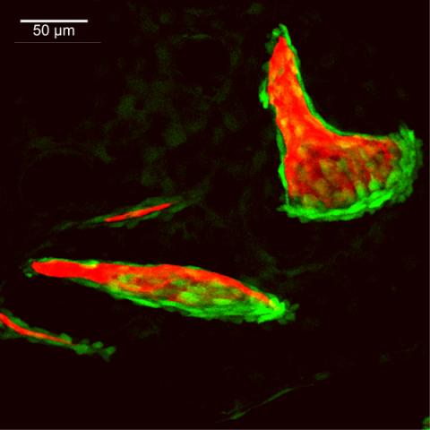
By 240 hpf, osteoblasts line the anterior edge of the opercle (op) as an additional growth mode begins to extend this edge anteriorly. Continued posteroventral outgrowth of the opercle adds substantial size to that seen at 192 hpf. The interopercle (iop) has expanded dorsal-ventrally along its posterior end as it grows toward the opercle. Ossification of the rod-shaped subopercle (sop) has initiated just ventral and medial to the opercle. Osteoblast populations of the opercle and subopercle are separated from one another by several cell layers of mesenchyme (data not shown). The branchiostegal rays are not visible in this particular plane of view. Bone matrix of the hyomandibula (hm) can be seen in the perichondrium of the hyosymplectic (not visible here), although the flattened, small osteoblasts of the hyomandibula are not seen at this magnification. Foci of EGFP-derived fluorescence across the image are derived from transgene expression in the ectoderm.
|
|
|
sp7:EGFP
Alizarin red
|
|
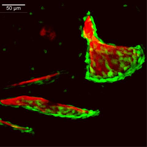
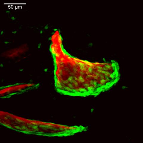
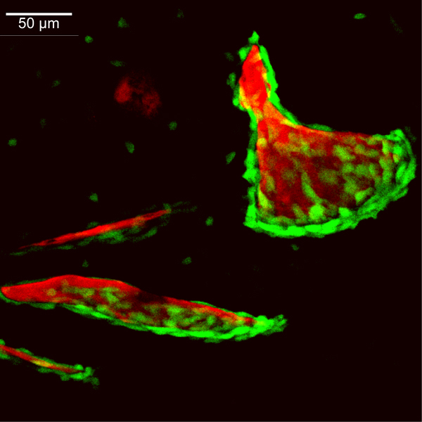
By 336 hpf, bone matrix has been added along the anterior edge of the opercle (op). The opercle, subopercle (sop), branchiostegal ray 3 (bsr3), and branchiostegal ray 2 (bsr2) are bordered by several cell layers of osteoblasts along their ventral and posterior edges, and primary outgrowth of these bones occurs in these regions. The ventral osteoblast population of the opercle overlaps laterally the dorsal edge of the subopercle. The posterior portion of the interopercle (iop) has continued to expand dorsal-ventrally and extend posteriorly to form a rounded end that overlaps laterally the anterior edge of the subopercle . The preopercle (pop) has also grown into the field of view just dorsal to the interopercle and overlaps laterally the hyomandibula (hm). The hyomandibula has extended a dermal projection from the perichondrium of the hyosymplectic (not visible here), and many osteoblasts are now visible along this bone. Foci of EGFP-derived fluorescence across the image are derived from transgene expression in the ectoderm.
|
|
|
sp7:EGFP
Alizarin red
|
|
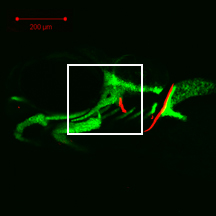
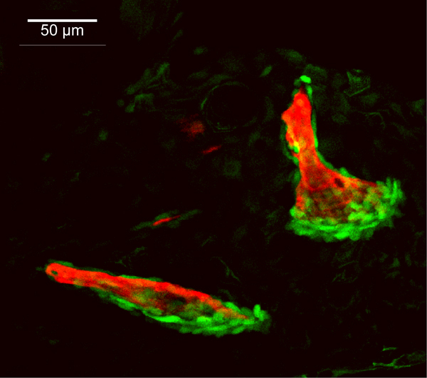
By 504hpf, the dermal bony skeleton, which is likely all derived from the second pharyngeal arch, covers completely the lateral aspect of the zebrafish head. The opercle (op) is trapezoidal in shape, due to the fact that a considerable amount of bone has been added along its dorsal and anterior edges, relative to that added along its ventral and posterior edges, since 336hpf. The anteroventral edge of the opercle articulates with the posterior edge of the interopercle (iop), overlapping it medially, and the ventral edge of the opercle articulates with the dorsal edge of the subopercle (sop), overlapping it laterally. The preopercle (pop) is now crescent-shaped with a posterodorsal extension along the anterior edge of the opercle. The ventral edge of the preopercle overlaps laterally the dorsal edge of the interopercle, and the dorsal edge of the preopercle overlaps laterally the hyomandibula (hm), which has extended a dermal projection from the perichondrium of the hyosymplectic (not visible here). The dorsal edge of branchiostegal ray 3 (bsr3) overlaps medially the ventral edge of the subopercle; the ventral edge of branchiosteal ray 3 overlaps laterally the dorsal edge of branchiostegal ray 2 (bsr2). The ventral edge of branchiostegal ray 2 overlaps laterally the dorsal edge of branchiostegal ray 1 (bsr1), which is the most anterior and latest-forming branchiostegal ray. The cleithrum (cl) of the shoulder girdle also is visible posteriorly in this particular view. |

 Movie(s):


