FishFace
21 dpf Development
| Info |
Image
Please hold mouse over images to see annotations.
|
|
|
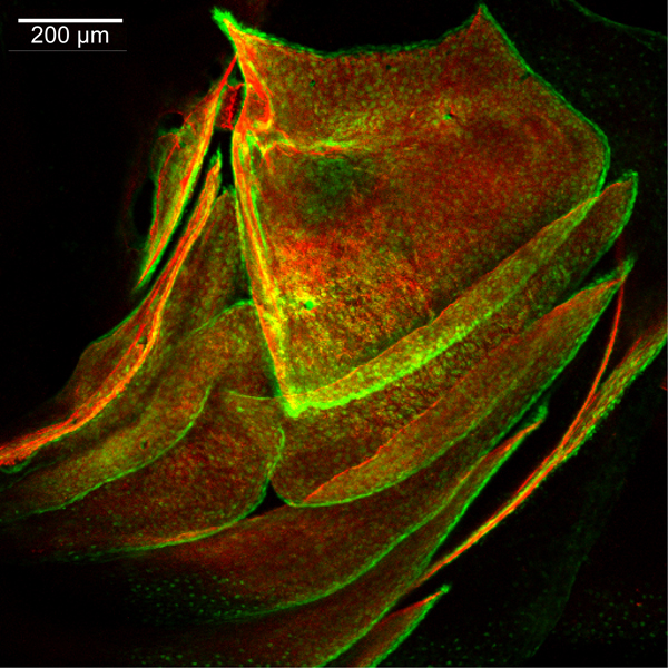
21 dpf
sp7:EGFP
Alizarin red
 |
|
|
| Description |
By 504hpf, the dermal bony skeleton, which is likely all derived from the second pharyngeal arch, covers completely the lateral aspect of the zebrafish head. The opercle (op) is trapezoidal in shape, due to the fact that a considerable amount of bone has been added along its dorsal and anterior edges, relative to that added along its ventral and posterior edges, since 336hpf. The anteroventral edge of the opercle articulates with the posterior edge of the interopercle (iop), overlapping it medially, and the ventral edge of the opercle articulates with the dorsal edge of the subopercle (sop), overlapping it laterally. The preopercle (pop) is now crescent-shaped with a posterodorsal extension along the anterior edge of the opercle. The ventral edge of the preopercle overlaps laterally the dorsal edge of the interopercle, and the dorsal edge of the preopercle overlaps laterally the hyomandibula (hm), which has extended a dermal projection from the perichondrium of the hyosymplectic (not visible here). The dorsal edge of branchiostegal ray 3 (bsr3) overlaps medially the ventral edge of the subopercle; the ventral edge of branchiosteal ray 3 overlaps laterally the dorsal edge of branchiostegal ray 2 (bsr2). The ventral edge of branchiostegal ray 2 overlaps laterally the dorsal edge of branchiostegal ray 1 (bsr1), which is the most anterior and latest-forming branchiostegal ray. The cleithrum (cl) of the shoulder girdle also is visible posteriorly in this particular view. |
 Movie(s):