FishFace
7 dpf Development
| Info |
Image
Please hold mouse over images to see annotations.
|
|
|
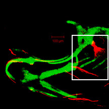
7 dpf
sp7:EGFP
Alizarin red
 |
|
|
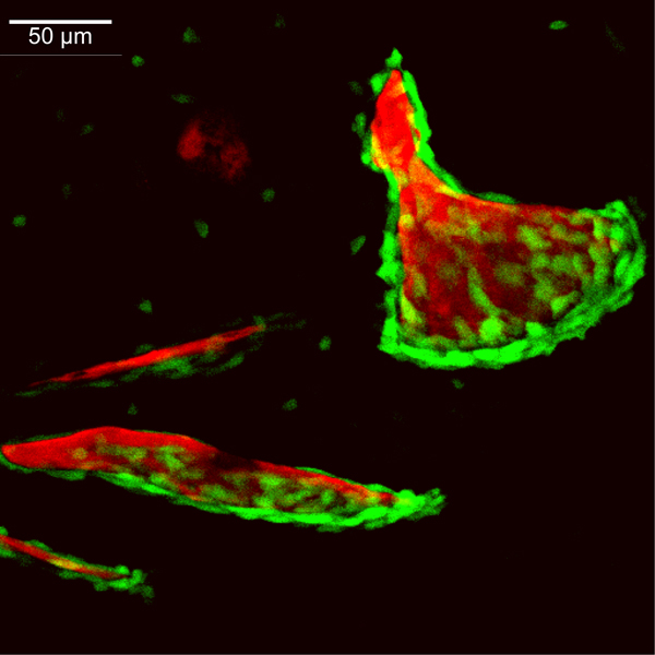
| Description | By 168 hpf, the ventral edge of the opercle (op) has continued its major mode of bone growth both ventrally and posteriorly, increasing in size from that seen at 144 hpf. Branchiostegal ray 3 (bsr3) continues to grow along its posteroventral edge. The interopercle (iop) has extended both anteriorly and posteriorly but remains a simple spicule of bone surrounded by a thin layer of osteoblasts. Likewise, branchiostegal ray 2 (bsr2) has remained rod-shaped, growing posteriorly, where numerous osteoblasts are visible. Bone matrix of the hyomandibula (hm) can be seen in the perichondrium of the hyosymplectic (not visible here), although the flattened, small osteoblasts of the hyomandibula are not seen at this magnification. Faint EGFP-derived fluorescence throughout the image is derived from transgene expression in the ectoderm. |
 Movie(s):