FishFace
8 dpf Development
| Info |
Image
Please hold mouse over images to see annotations.
|
|
|
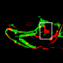
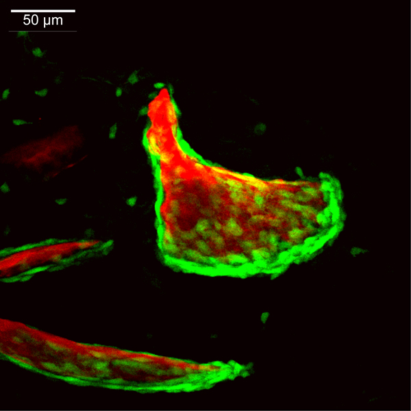
8 dpf
sp7:EGFP
Alizarin red
 |
|
|
| Description | By 192 hpf, continued posteroventral expansion of the opercle (op) and branchiostegal ray 3 (bsr3) results in overall size increases in these bones relative to that seen at 168 hpf. The interopercle (iop) has grown in dorsal-ventral width and has more osteoblasts along its edges than seen at 168 hpf. Bone matrix of the hyomandibula (hm) can be seen in the perichondrium of the hyosymplectic (not visible here), although the flattened, small osteoblasts of the hyomandibula are not seen at this magnification. Foci of EGFP-derived fluorescence across the image are derived from transgene expression in the ectoderm. |
 Movie(s):