FishFace
14 dpf Development
| Info |
Image
Please hold mouse over images to see annotations.
|
|
|
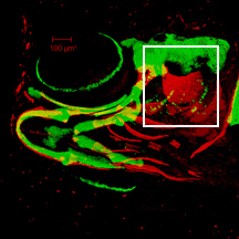
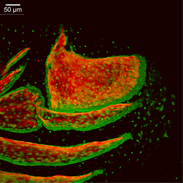
14 dpf
sp7:EGFP
Alizarin red
 |
|
|
| Description | By 336 hpf, bone matrix has been added along the anterior edge of the opercle (op). The opercle, subopercle (sop), branchiostegal ray 3 (bsr3), and branchiostegal ray 2 (bsr2) are bordered by several cell layers of osteoblasts along their ventral and posterior edges, and primary outgrowth of these bones occurs in these regions. The ventral osteoblast population of the opercle overlaps laterally the dorsal edge of the subopercle. The posterior portion of the interopercle (iop) has continued to expand dorsal-ventrally and extend posteriorly to form a rounded end that overlaps laterally the anterior edge of the subopercle . The preopercle (pop) has also grown into the field of view just dorsal to the interopercle and overlaps laterally the hyomandibula (hm). The hyomandibula has extended a dermal projection from the perichondrium of the hyosymplectic (not visible here), and many osteoblasts are now visible along this bone. Foci of EGFP-derived fluorescence across the image are derived from transgene expression in the ectoderm. |
 Movie(s):