FishFace
6 dpf Development
| Info |
Image
Please hold mouse over images to see annotations.
|
|
|
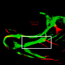
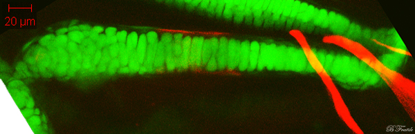
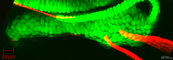
6 dpf
zc81Tg
Alizarin red
 |
|
|
| Description |
By 150 hpf, the ceratohyal (ch) has grown considerably from that seen at 104 hpf. The region consisting of one cell layer of chondrocytes in the mediolateral dimension now extends to the posterior end, suggesting that cell intercalation continues to drive ceratohyal growth in length. Clusters of chondrocytes at both ends of the ceratohyal in the lateral view gives the overall morphology the appearance of a dumbbell. The ceratohyal bone (chb) can be seen in the ceratohyal perichondrium at the middle region, while the ventral hypohyal (hhv) can be seen in some specimens in the perichondrium at the ventro-anterior tip. The ceratohyal continues to articulate with the interhyal (ih) along the dorso-posterior edge of the ceratohyal, with the basihyal (bh) and contralateral ceratohyal at the anterior midline, and with branchiostegal ray 3 (bsr3) along the postero-lateral aspect of the ceratohyal. More anterior to branchiostegal ray 3, branchiostegal ray 2 (bsr2) projects postero-ventrally from the postero-lateral aspect of the ceratohyal. |


 Movie(s):