FishFace
36 hpf Development
[ Back to Pharyngeal development: Pre-skeletal]
| Info | Image Please hold mouse over images to see annotations. |
|
|
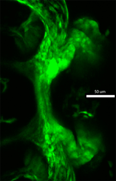
36 hpf
fli1a:EGFP
|
|
|
| Description |
By 36 hpf, pharyngeal arch 1 (pa1) appears much longer than pharyngeal arch 2 (pa2) along the dorsal-ventral axis. Pharyngeal pouch 1 (pp1), which is GFP-negative in fli1a:EGFP transgenic fish, continues to separate pharyngeal arches 1 and 2 dorsally, while the oral ectoderm (oe), which also is GFP-negative in fli1a:EGFP transgenic fish, continues to separate dorsal and ventral cells in the anterior region of pharyngeal arch 1. Neural crest cells in the posterodorsal region of pharyngeal arch 1 and in the anterodorsal region of pharyngeal arch 2 appear to be aligned with the surface of pharyngeal pouch 1. A ventral view demonstrates that the ventral region of pharyngeal arch 1 has begun to extend medially. |