FishFace
48 hpf Development
[ Back to Pharyngeal development: Pre-skeletal]
| Info | Image Please hold mouse over images to see annotations. |
|
|
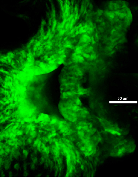
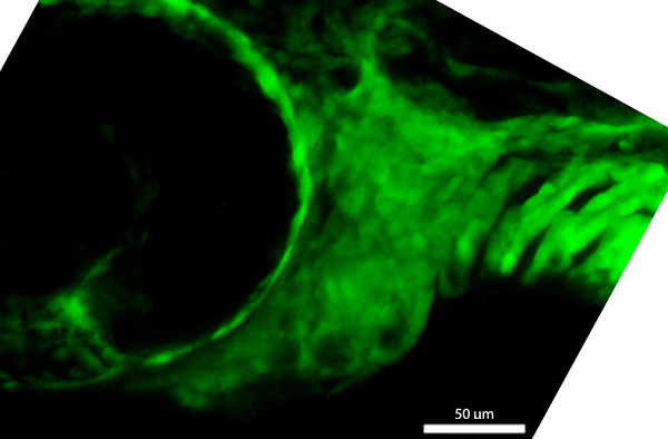
48 hpf
fli1a:EGFP
|
|
|
| Description |
By 48 hpf, the ventral portions of pharyngeal arches 1 and 2 (pa1, pa2) have no clear boundary between them, and the dorsal portion of pharyngeal arch 1 is no longer visible from lateral view. The posterior pharyngeal arches 3-6 (pa3-6), which contribute cells to the ceratobranchials of the pharyngeal skeleton, are visible in a series with similar morphology. The ventral region of pharyngeal arch 1 has expanded extensively from that seen at 42 hpf, so that cells from the left and right sides of the embryo meet at the midline. In addition, the oral ectoderm (oe), which is GFP-negative in fli1a:EGFP transgenic fish, is more constricted medially. Compared to that seen at 42hpf, the ventral region of pharyngeal arch 2 has extended further medially, but cells of the left and right sides have yet to meet at the midline. |