User Guide & Data Access
User Guide
A user guide to facilitate navigation in EarBase is under development and will be released soon. A link will appear here to download the PDF file. Updates will be made periodically.
Download User Guide Coming soonData Access
Access to EarBase content is divided into two tiers: Open and Controlled. Open Access permits unregistered visitors to browse content, but downloads are restricted. Approval for Controlled Access requires application to and review by the Data Access Committee. This is necessary for compliance with privacy laws and NIH guidelines.
Review the procedures listed under the POLICIES tab on the navbar above for detailed background and instructions.
Download Step-by-Step DAC GuideContent Availability: Overview
By default, visitors to EarBase (and all FaceBase consortia) are viewing content at the Open Access level. Visitors may search and browse Cases and Specimens using keywords. Downloading content is limited. The available content should be sufficient to determine whether Controlled Access privileges will facilitate the proposed research.
Users approved for Controlled Access can access additional details about Cases and Specimens, view and download full resolution images, and download approved content.
Open Access
Available to all visitors
- Browse and search Cases and Specimens
- View otologic diagnoses (summary)
- View histopathology descriptions
- View specimen information and assay listings
- View image thumbnails
- View case summary information
Controlled Access
Requires DAC approval
- All Open Access content, plus:
- Full otologic history
- Full resolution images
- Downloads (as designated during approval)
- Diagnostics, summaries, images, analyses
Examples of Open and Controlled Content
The following examples are drawn from a single Case to illustrate the difference between access levels.
Open Access Level (examples)
Otologic diagnoses (summary)
- Mastoidectomy, modified radical, left.
- Fibrocystic sclerosis, peripheral cells of the mastoid.
- Ulceration, walls of the mastoid bowl.
- Atrophy, organ of Corti, left.
- Normal appearing facial nerve.
- Stria vascularis, atrophy, mild, right.
Histopathology
- Resorption of the head of the malleus and part of the manubrium. The body of the incus is intact but part of the long process is resorbed. The stapes is intact and in its normal position. The tympanic membrane is greatly thickened.
- In the basal 10 mm of the cochlea, the organ of Corti is missing. Elsewhere it is present but contains only a few scattered hair cells, possibly only 10 to 20% remaining.
- The stria vascularis is severely atrophied throughout the basal and middle turns.
- The macula sacculi appears to have a flattened sensory epithelium and some loss of hair cells. The cristae and the utricular macula appear to contain hair cells but possibly some are missing.
- Only 10 to 20% of the cochlear neurons in the basal turn, about 70% in the middle turn, and about 90% in the apical turn.
Specimen info
Listings of tissues acquired, assays conducted, image thumbnails.
Controlled Access Level (examples of additional content available)
Full otologic history
The patient had a left mastoidectomy performed in childhood and suffered a profound hearing loss and a total left facial paralysis. It is not known whether these functional losses were caused by the infection or by the surgery. At the age of 77 he complained of pain and swelling behind the left ear. An audiogram revealed a profound hearing loss in the left ear and mild sensorineural hearing loss characterized by a flat audiometric pattern in the right ear. Speech discrimination was 88% on the right and zero on the left. Caloric tests were not performed. An operative procedure was performed to drain an abscess of the mastoid area and to enlarge the external auditory meatus. This resulted in a dry asymptomatic ear. Examination of the mastoid area at the time of this surgery revealed the frayed ends of the facial nerve in its descending portion and was interpreted to be the result of previous surgical injury. The middle ear area appeared uninfected.
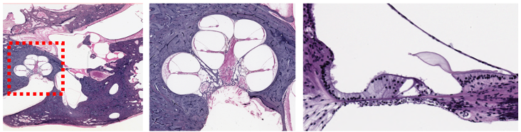
Full resolution images
High-resolution scans available for download as approved.
Downloads
Diagnostics, summaries, images, and analyses as designated during the approval process.
Prior to their release, patient (Case) records were scrubbed to remove Protected Health Information (PHI) and Personally Identifiable Information (PII), following HIPAA and NIH guidelines (Requirements for NIH Controlled-Access Data Repositories and Users). Also redacted or revised were instances of inappropriate and outdated terms. Please contact us if you discover questionable content in EarBase.