FishFace
Pharyngeal development - Skeletal
This series of images from late embryonic stages--2 days post-fertilization (dpf)--to late larval stages—21 dpf--shows the appearance, overall anatomical arrangements, and gross morphology of cartilage and bone elements of the developing zebrafish craniofacial apparatus. We use the zc81Tg transgenic zebrafish, with fluorescent chondrocytes, to visualize cartilage, and Alizarin red to visualize bone matrix. Images are taken from both ventral and lateral views.
| Info
Click linked text in this column to see full page image(s)
|
Image
Please hold mouse over images to see annotations.
Click on magnifying icon to view large image. |
Description
Click on an underlined term to see its definition and link to related pages
|
|
|
zc81Tg
|
|
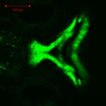
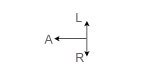
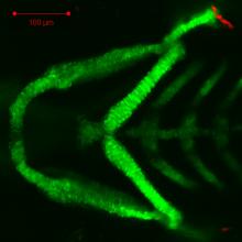
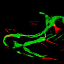
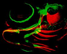
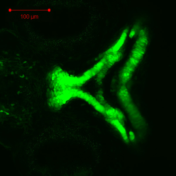
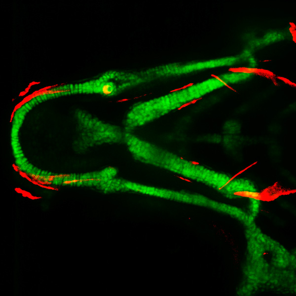
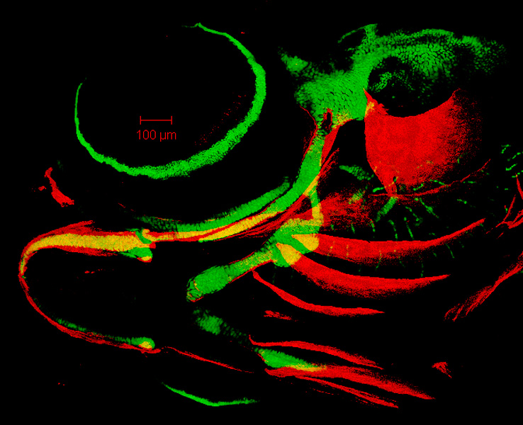
By 55 hpf, developing cartilage cells of the craniofacial skeleton are apparent in discrete cartilage elements, such as the trabecula (t) and the ethmoid (e) of the neurocranium, Meckel’s cartilage (Mk); and the palatoquadrate (pq), which are derived from the first pharyngeal arch, and the ceratohyal (ch) and the hyosymplectic (hs), which are derived from the second pharyngeal arch. All of these cartilage cells are surrounded by a secreted cartilage matrix at this timepoint (data not shown). |
|
|
zc81Tg
Alizarin red
|
|
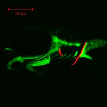
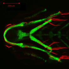
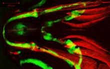
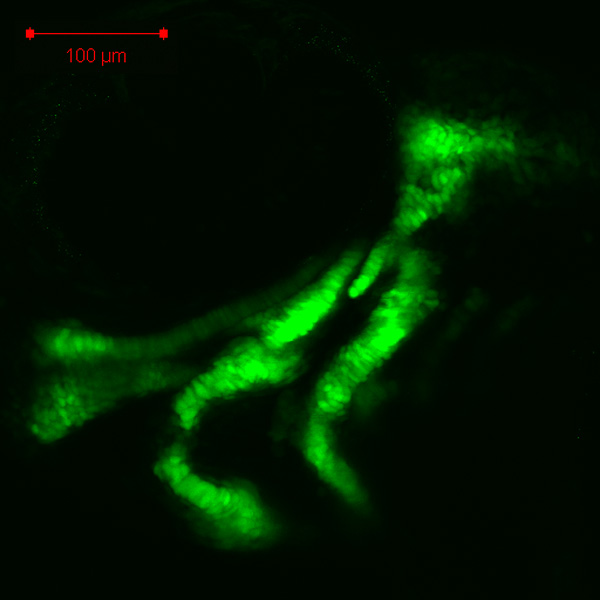
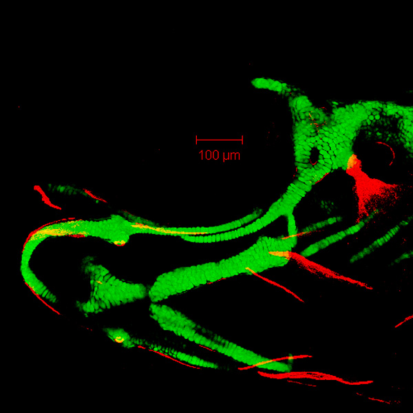
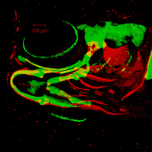
By 78 hpf, developing cartilages of the pharyngeal skeleton, such as Meckel’s cartilage (Mk), the palatoquadrate (pq), and the ceratohyal (ch), have grown dramatically, compared to 55 hpf. Also visible are the basihyal (bh), hyosymplectic (hs), ceratobranchial (cb), copula 1 (cop1), and hypobranchial (hb) cartilages. Alizarin red staining indicates the appearance of the opercle (op) bone, derived from the second pharyngeal arch near the dorsal part of the hyosymplectic, during the third day of development. Also visible are the cleithrum (cl) bone and the coraco-scapular (cor) cartilage of the pectoral fin skeleton. |
|
|
zc81Tg
Alizarin red
|
|
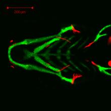
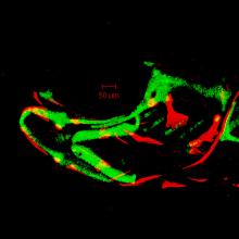
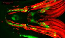
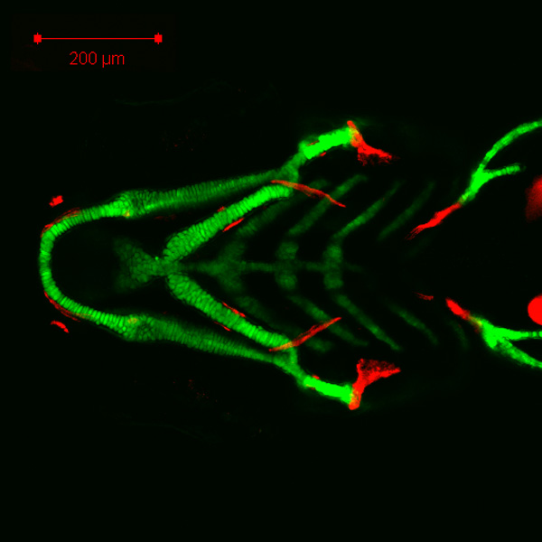
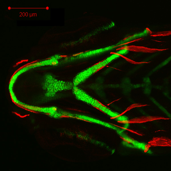
By 105 hpf, this decreased magnification view shows the basic anatomical arrangement of the entire early zebrafish pharyngeal skeleton, including Meckel’s cartilage (Mk), palatoquadrate (pq), basihyal (bh), ceratohyal (ch), interhyal (ih), hyosymplectic (c) (hs), copula 1, and hypobranchial cartilages and the ceratobranchial bone (cbb). Alizarin red staining indicates the opercle (op) and ceratobranchial 5 (cb5) bones, as well as the appearance of the maxilla (mx), dentary (d), and retroarticular (ra) bones, derived from the first pharyngeal arch, and ceratohyal bone (chb), branchiostegal ray 3 (bsr3), and hyomandibula (hm) bones, derived from the second pharyngeal arch, during the fourth day of development. Also visible are the cleithrum (cl) bone and the coraco-scapular (cor) cartilage of the pectoral fin skeleton. |
|
|
zc81Tg
Alizarin red
|
|
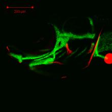
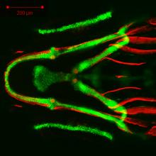
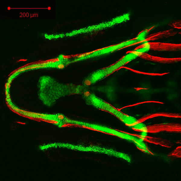
By 151 hpf, the pharyngeal skeleton includes Meckel’s (Mk), palatoquadrate (pq), basihyal (bh), ceratohyal (ch), interhyal (ih), hyosymplectic (hs), ceratobranchial (cb), copula 1, and hypobranchial cartilages. Alizarin red staining indicates the maxilla (mx), dentary (d), retroarticular (ra), entopterygoid (en), ceratohyal bone (chb), branchiostegal ray 3 (bsr3), opercle (op), and hyomandibula (hm) bones, as well as the appearance of the branchiostegal ray 2 (bsr2) bone, derived from the second pharyngeal arch. |
|
|
zc81Tg
Alizarin red
|
|
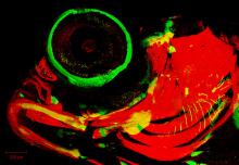
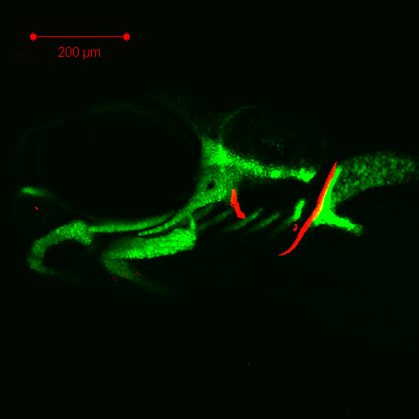
By 199 hpf, the pharyngeal skeleton includes Meckel’s (Mk), palatoquadrate (pq), basihyal (bh), ceratohyal (ch), interhyal (ih), hyosymplectic (hs), ceratobranchial (cb), copula 1, and hypobranchial cartilages, and maxilla (mx), dentary (d), retroarticular (ra), entopterygoid (en), ceratohyal bone (chb), hyomandibula (hm), opercle (op), and ceratobranchial 5 (cb5) bones. The branchiostegal rays now comprise the initial two posterior (bsr2, 3), as well as one more anterior (bsr1), bones. Other bones visible here between 6 dpf and 8 dpf include the anguloarticular (aa) and quadrate (q) bones, derived from the first pharyngeal arch, and the interopercle (iop) and subopercle (sop) bones, derived from the second pharyngeal arch. Also visible are the scleral (sc) cartilage of the eye and the coraco-scapular (cor) cartilage and the cleithrum (cl) bone of the pectoral fin skeleton. |
|
|
zc81Tg
Alizarin red
|
|
By 247 hpf, the pharyngeal skeleton includes Meckel’s (Mk), palatoquadrate (pq), basihyal (bh), ceratohyal (ch), interhyal (ih), and hyosymplectic (hs) cartilages, and maxilla (mx), dentary (d), anguloarticular (aa), retroarticular (ra), quadrate (q), ceratohyal bone (chb), branchiostegal rays 1-3 (bsr1, 2, 3), hyomandibula (hm), interopercle (iop), subopercle (sop), opercle (op), and ceratobranchial 5 (cb5) bones. Other bones visible here between 8 dpf and 10 dpf include the premaxilla (pm), derived from the first pharyngeal arch, the basihyal (bhb), ventral hypohyal (hhv), symplectic (sy), and preopercle (pop) bones, derived from the second pharyngeal arch, and the urohyal (uh) bone. Also visible are the scleral (sc) cartilage of the eye and the cleithrum (cl) bone of the pectoral fin skeleton. |
|
|
zc81Tg
Alizarin red
|
|
By 340 hpf, developing elements of the pharyngeal skeleton have grown dramatically, compared to 247 hpf. Elements shown here include Meckel’s (Mk), palatoquadrate (pq), basihyal (bh), ceratohyal (ch), interhyal (ih), and hyosymplectic (hs) cartilages, and premaxilla (pm), maxilla (mx), dentary (d), anguloarticular (aa), retroarticular (ra), quadrate (q), ventral hypohyal (hhv), ceratohyal bone (chb), branchiostegal rays 1-3 (bsr1, 2, 3), symplectic (sy), hyomandibula (hm), preopercle (pop), interopercle (iop), subopercle (sop), opercle (op), urohyal (uh), and ceratobranchial 5 (cb5) bones. Also visible are the scleral (sc) cartilage of the eye and the cleithrum (cl) bone of the pectoral fin skeleton. |
|
|
zc81Tg
Alizarin red
|
|
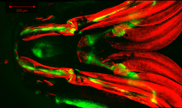
By 514 hpf, growth of the bony elements of the pharyngeal skeleton have almost completely engulfed cartilage elements and also expanded across the ventral portion of the gills. Elements shown here include Meckel’s (Mk), palatoquadrate (pq), basihyal (bh), ceratohyal (ch), interhyal (ih), and hyosymplectic (hs) cartilages, and premaxilla (pm), maxilla (mx), dentary (d), anguloarticular (aa), retroarticular (ra), quadrate (q), ventral hypohyal (hhv), ceratohyal bone (chb), branchiostegal rays 1-3 (bsr1, 2, 3), urohyal (uh), symplectic (sy), preopercle (pop), interopercle (iop), subopercle (sop), and opercle (op) bones. Becoming visible here between 14 dpf and 21 dpf are the infraorbital 1 (inf1) and metapterygoid (mpt) bones, likely derived from the first pharyngeal arch, and the epihyal (eh) bone, derived from the second pharyngeal arch. Also visible are the scleral (sc) cartilage of the eye and the cleithrum (cl) bone of the pectoral fin skeleton. |
 Movie(s):

